| CONDENSED MATTER: STRUCTURAL, MECHANICAL, AND THERMAL PROPERTIES |
Prev
Next
|
|
|
Characterization of swift heavy ion tracks in MoS2 by transmission electron microscopy |
| Li-Jun Xu(徐丽君)1,2, Peng-Fei Zhai(翟鹏飞)1,2,†, Sheng-Xia Zhang(张胜霞)1, Jian Zeng(曾健)1,2, Pei-Pei Hu(胡培培)1, Zong-Zhen Li(李宗臻)1,2, Li Liu(刘丽)1,2, You-Mei Sun(孙友梅)1,2, and Jie Liu(刘杰)1,2,‡ |
1 Institute of Modern Physics, Chinese Academy of Sciences, Lanzhou 730000, China
2 School of Nuclear Science and Technology, University of Chinese Academy of Sciences, Beijing 100049, China |
|
|
|
|
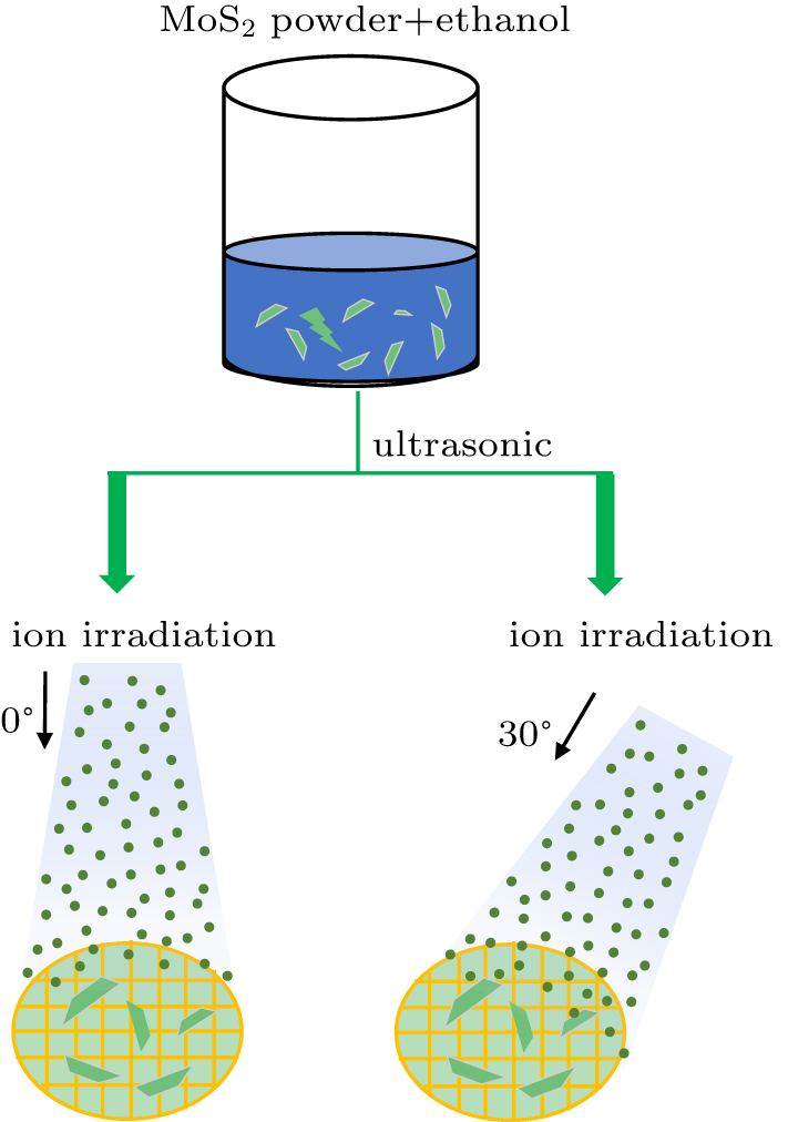
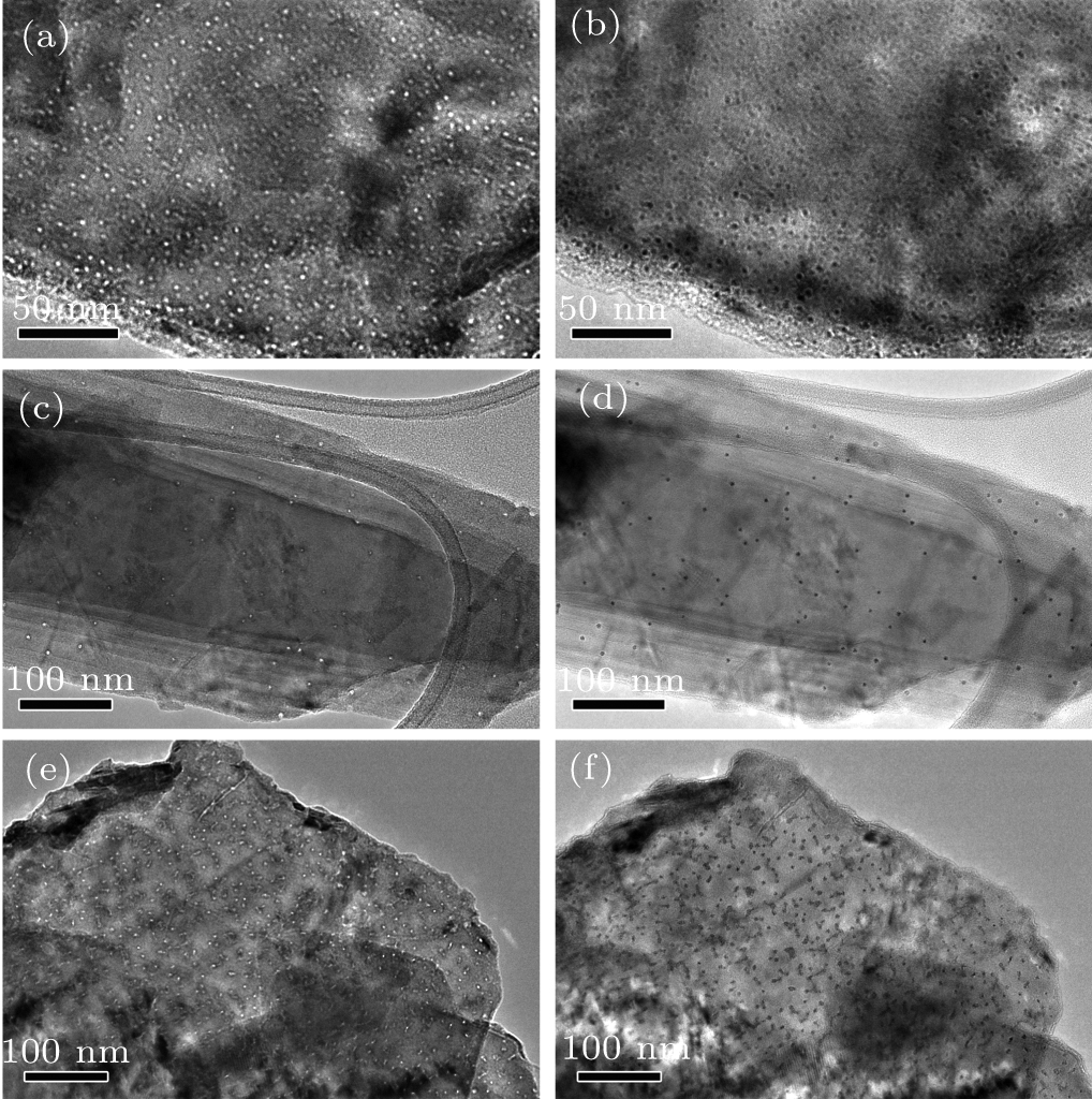
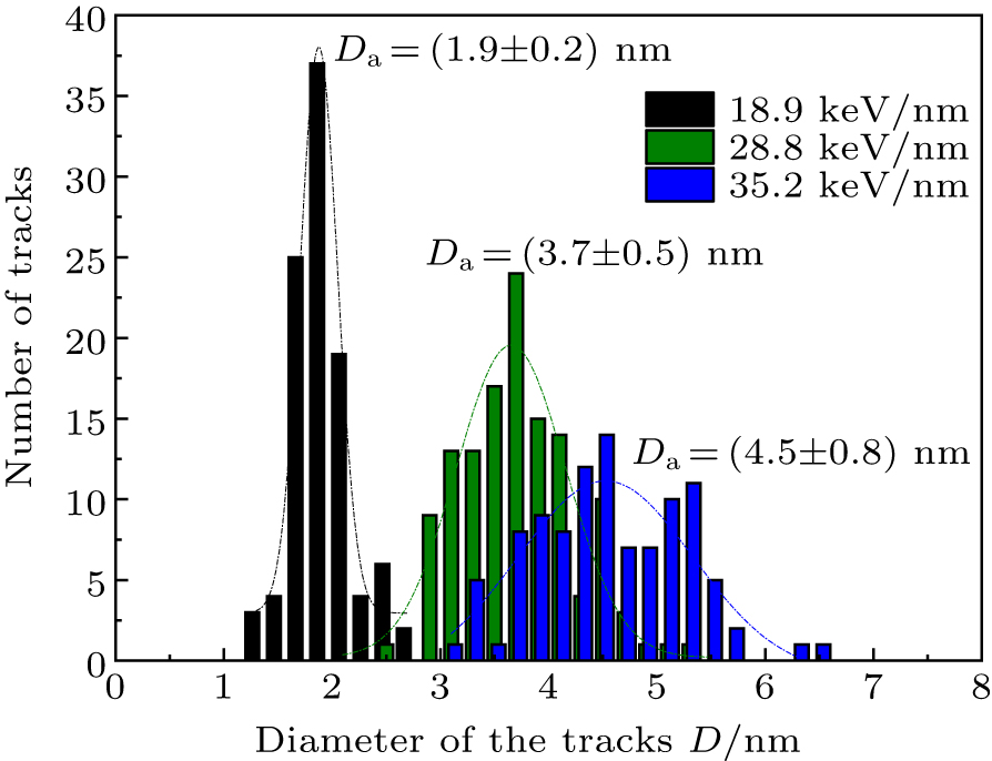
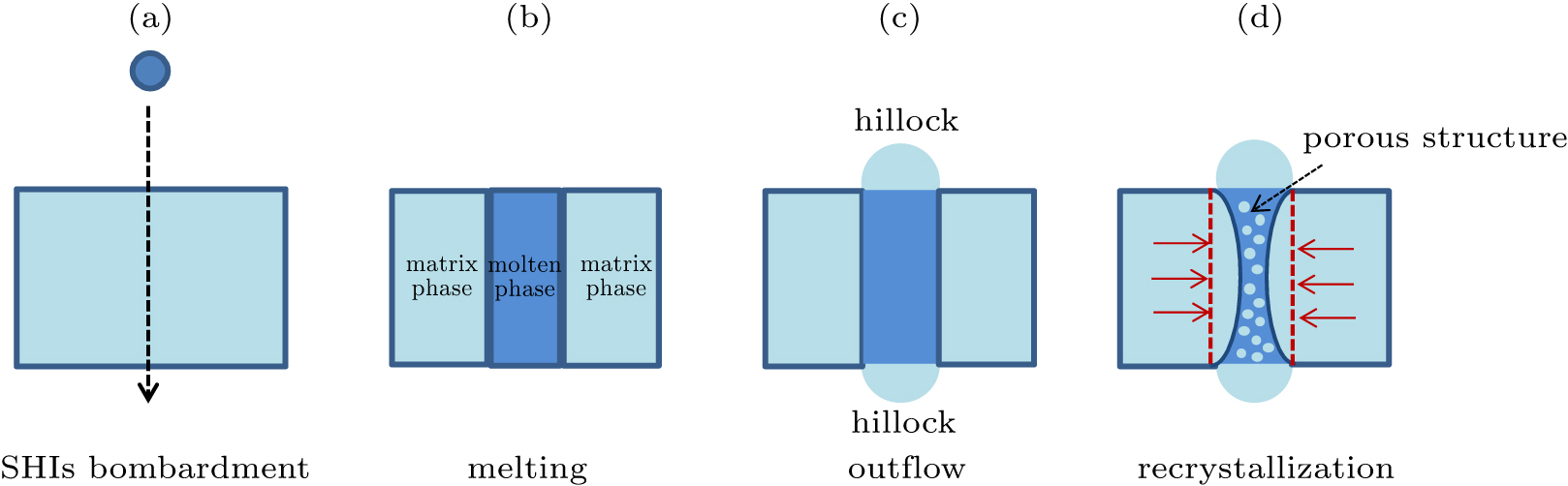
Abstract The various morphologies of tracks in MoS2 irradiated by swift heavy ions at normal and 30° incidence with 9.5–25.0 MeV/u 86Kr, 129Xe, 181Ta, and 209Bi ions were investigated by transmission electron microscopy. The diameter of ion tracks increases from 1.9 nm to 4.5 nm with increasing electronic energy loss. The energy loss threshold of the track formation in MoS2 is predicted as about 9.7 keV/nm based on the thermal spike model and it seems consistent with the experimental results. It is shown that the morphology of ion tracks is related to the penetration length of ions in MoS2. The formation process of ion tracks is discussed based on the cooperative process of outflow and recrystallization of the molten phase during rapid quenching.
|
Received: 27 April 2020
Revised: 06 August 2020
Accepted manuscript online: 07 August 2020
|
|
PACS:
|
61.80.-x
|
(Physical radiation effects, radiation damage)
|
| |
61.82.-d
|
(Radiation effects on specific materials)
|
| |
68.37.Lp
|
(Transmission electron microscopy (TEM))
|
|
|
Corresponding Authors:
†Corresponding author. E-mail: zhaipengfei@impcas.ac.cn ‡Corresponding author. E-mail: j.liu@impcas.ac.cn
|
| About author: †Corresponding author. E-mail: zhaipengfei@impcas.ac.cn ‡Corresponding author. E-mail: j.liu@impcas.ac.cn * Project supported by the National Natural Science Foundation of China (Grant Nos. 11675233, 11690041, 11405229, 11705246, and 11505243), Chinese Academy of Sciences “Light of West China” Program, and the Youth Innovation Promotion Association of Chinese Academy of Sciences (Grant No. 2020412). |
Cite this article:
Li-Jun Xu(徐丽君), Peng-Fei Zhai(翟鹏飞)†, Sheng-Xia Zhang(张胜霞), Jian Zeng(曾健), Pei-Pei Hu(胡培培), Zong-Zhen Li(李宗臻), Li Liu(刘丽), You-Mei Sun(孙友梅), and Jie Liu(刘杰)‡ Characterization of swift heavy ion tracks in MoS2 by transmission electron microscopy 2020 Chin. Phys. B 29 106103
|
| [1] |
Dufour Ch, Audouard A, Beneu F, Dural J, Girard J P, Hairie A, Levalois M, Paumier E, Toulemonde M 1993 J. Phys.: Condens. Matter 5 4573 DOI: 10.1088/0953-8984/5/26/027 |
| [2] |
|
| [3] |
|
| [4] |
Mara A, Siwy Z, Trautmann C, Wan J, Kamme F 2004 Nano Lett. 4 497 DOI: 10.1021/nl035141o |
| [5] |
Fink D, Chadderton L T, Kiv A, Saad A, Tabacnics M, Rizutto M, de A, Silva A, de O D, Fahrner W R, Hoppe K 2007 Radiat. Eff. Defects Solids 162 543 DOI: 10.1080/10420150701470746 |
| [6] |
Madauß L, Zegkinoglou I, Muiños H V, Choi Y W, Kunze S, Zhao M Q, Naylor C H, Ernst P, Pollmann E, Ochedowski O, Lebius H, Benyagoub A, Ban-d’Etat B, Johnson A T C, Djurabekova F, Cuenya B R, Schleberger M 2018 Nanoscale 10 22908 DOI: 10.1039/C8NR04696D |
| [7] |
|
| [8] |
Vazquez H, Åhlgren E H, Ochedowski O, Leino A A, Mirzayev R, Kozubek R, Lebius H, Karlusic M, Jaksic M, Krasheninnikov A V, Kotakoski J, Schleberger M, Nordlund K, Djurabekova F 2017 Carbon 114 511 DOI: 10.1016/j.carbon.2016.12.015 |
| [9] |
Akcöltekin S, Bukowska H, Peters T, Osmani O, Monnet I, Alzaher I, Ban d’Etat B, Lebius H, Schleberger M 2011 Appl. Phys. Lett. 98 103103 DOI: 10.1063/1.3559619 |
| [10] |
|
| [11] |
Madauß L, Ochedowski O, Lebius H, Ban-d’Etat B, Naylor C H, Johnson A T C, Kotakoski J, Schleberger M 2017 2D Mater. 4 015034 DOI: 10.1088/2053-1583/4/1/015034 |
| [12] |
Radisavljevic B, Radenovic A, Brivio J, Giacometti V, Kis A 2011 Nat. Nanotechnol. 6 147 DOI: 10.1038/nnano.2010.279 |
| [13] |
|
| [14] |
|
| [15] |
Kang Y M, Najmaei S, Liu Z, Bao Y J, Wang Y M, Zhu X, Halas N J, Nordlander P, Ajayan P M, Lou J, Fang Z Y 2014 Adv. Mater. 26 6467 DOI: 10.1002/adma.201401802 |
| [16] |
|
| [17] |
Zhai P F, Nan S, Xu L J, Li W X, Li Z Z, Hu P P, Zeng J, Zhang S X, Sun Y M, Liu J 2019 Nucl. Instr. Meth. Phys. Res. Sect. B 457 72 DOI: 10.1016/j.nimb.2019.07.024 |
| [18] |
O’Connell J, Skuratov V, van Vuuren A J, Saifulin M, Akilbekov A 2016 Phys. Status Solidi B 253 2144 DOI: 10.1002/pssb.201600473 |
| [19] |
Garrido F, Moll S, Sattonnay G, Thomé L, Vincent L 2009 Nucl. Instr. Meth. Phys. Res. Sect. B 267 1451 DOI: 10.1016/j.nimb.2009.01.070 |
| [20] |
Lang M, Toulemonde M, Zhang J, Zhang F, Tracy C L, Lian J, Wang Z, Weber W J, Severin D, Bender M, Trautmann C, Ewing R C 2014 Nucl. Instr. Meth. Phys. Res. Sect. B 336 102 DOI: 10.1016/j.nimb.2014.06.019 |
| [21] |
|
| [22] |
Schattat B, Bolse W, Klaumünzer S, Zizak I, Scholz R 2005 Appl. Phys. Lett. 87 173110 DOI: 10.1063/1.2115084 |
| [23] |
|
| [24] |
|
| [25] |
Chase M W 1998 Journal of Physical Chemical Reference Data Monograph No. 9 New York American Institute of Physics 1594
|
| [26] |
|
| [27] |
|
| [28] |
|
| [29] |
Toulemonde M, Assmann W, Dufour C, Meftah A, Studer F, Trautmann C 2006 Symposium on Ion Beam Science - Solved and Unsolved Problems May 1–5, 2006 Copenhagen, Denmark 263
|
| [30] |
Meftah A, Brisard F, Costantini J, Hage-Ali M, Stoquert J, Studer F, Toulemonde M 1993 Phys. Rev. B 48 920 DOI: 10.1103/PhysRevB.48.920 |
| [31] |
Zhai P F, Liu J, Duan J L, Chang H L, Zeng J, Hou M D, Sun Y M 2011 Nucl. Instr. Meth. Phys. Res. Sect. B 269 2035 DOI: 10.1016/j.nimb.2011.06.010 |
| [32] |
|
| [33] |
|
| [34] |
|
| [35] |
Li W, Kluth P, Schauries D, Rodriguez M, Lang M, Zhang F, Zdorovets M, Trautmann C, Ewing R C 2014 Am. Mineral. 99 1127 DOI: 10.2138/am.2014.4669 |
| [36] |
|
| [37] |
|
| [38] |
Rymzhanov R A, Medvedev N, O’Connell J H, Janse van Vuuren A, Skuratov V A, Volkov A E 2019 Sci. Rep. 9 3837 DOI: 10.1038/s41598-019-40239-9 |
| [39] |
Petkov V, Billinge S J L, Larson P, Mahanti S D, Vogt T, Rangan K K, Kanatzidis M G 2002 Phys. Rev. B 65 092105 DOI: 10.1103/PhysRevB.65.092105 |
| [40] |
Jang J T, Jeong S, Seo J W, Kim M C, Sim E, Oh Y, Nam S, Park B, Cheon J 2011 J. Am. Chem. Soc. 133 7636 DOI: 10.1021/ja200400n |
| [41] |
Manna L, Wang L W, Cingolani R, Alivisatos A P 2005 J. Phys. Chem. B 109 6183 DOI: 10.1021/jp0445573 |
| [42] |
Xue X X, Feng Y X, Chen K Q, Zhang L X 2018 J. Chem. Phys. 148 134704 DOI: 10.1063/1.5010996 |
| [43] |
|
| No Suggested Reading articles found! |
|
|
Viewed |
|
|
|
Full text
|
|
|
|
|
Abstract
|
|
|
|
|
Cited |
|
|
|
|
Altmetric
|
|
blogs
Facebook pages
Wikipedia page
Google+ users
|
Online attention
Altmetric calculates a score based on the online attention an article receives. Each coloured thread in the circle represents a different type of online attention. The number in the centre is the Altmetric score. Social media and mainstream news media are the main sources that calculate the score. Reference managers such as Mendeley are also tracked but do not contribute to the score. Older articles often score higher because they have had more time to get noticed. To account for this, Altmetric has included the context data for other articles of a similar age.
View more on Altmetrics
|
|
|