|
Special Issue:
SPECIAL TOPIC — Modeling and simulations for the structures and functions of proteins and nucleic acids
|
| TOPICAL REVIEW—Modeling and simulations for the structures and functions of proteins and nucleic acids |
Prev
Next
|
|
|
Structural and dynamical mechanisms of a naturally occurring variant of the human prion protein in preventing prion conversion |
| Yiming Tang(唐一鸣), Yifei Yao(姚逸飞), and Guanghong Wei(韦广红)† |
| 1 Department of Physics, State Key Laboratory of Surface Physics and Key Laboratory for Computational Physical Science (Ministry of Education), and Multiscale Research Institute of Complex Systems, Fudan University, Shanghai 200433, China |
|
|
|
|
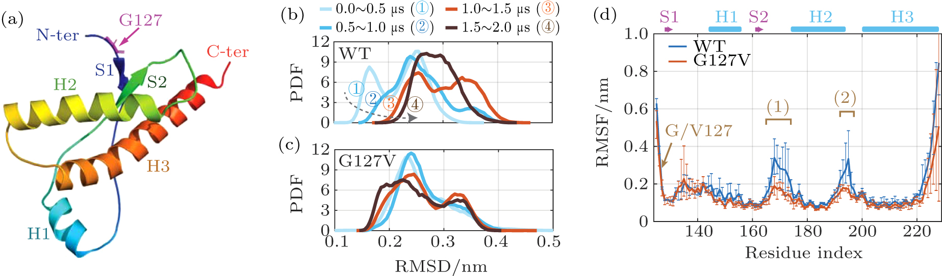
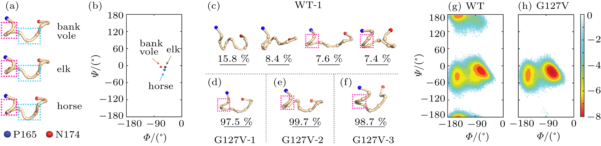
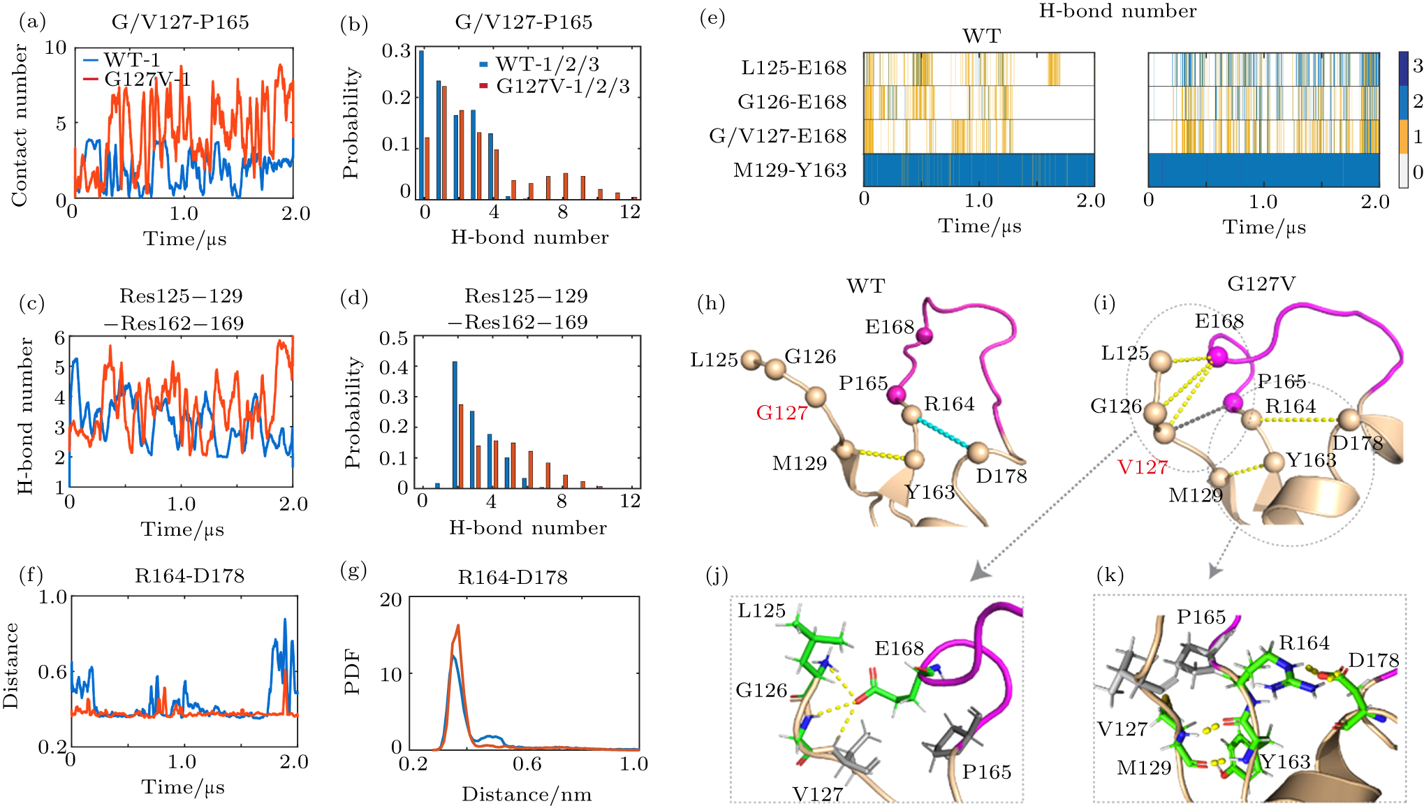
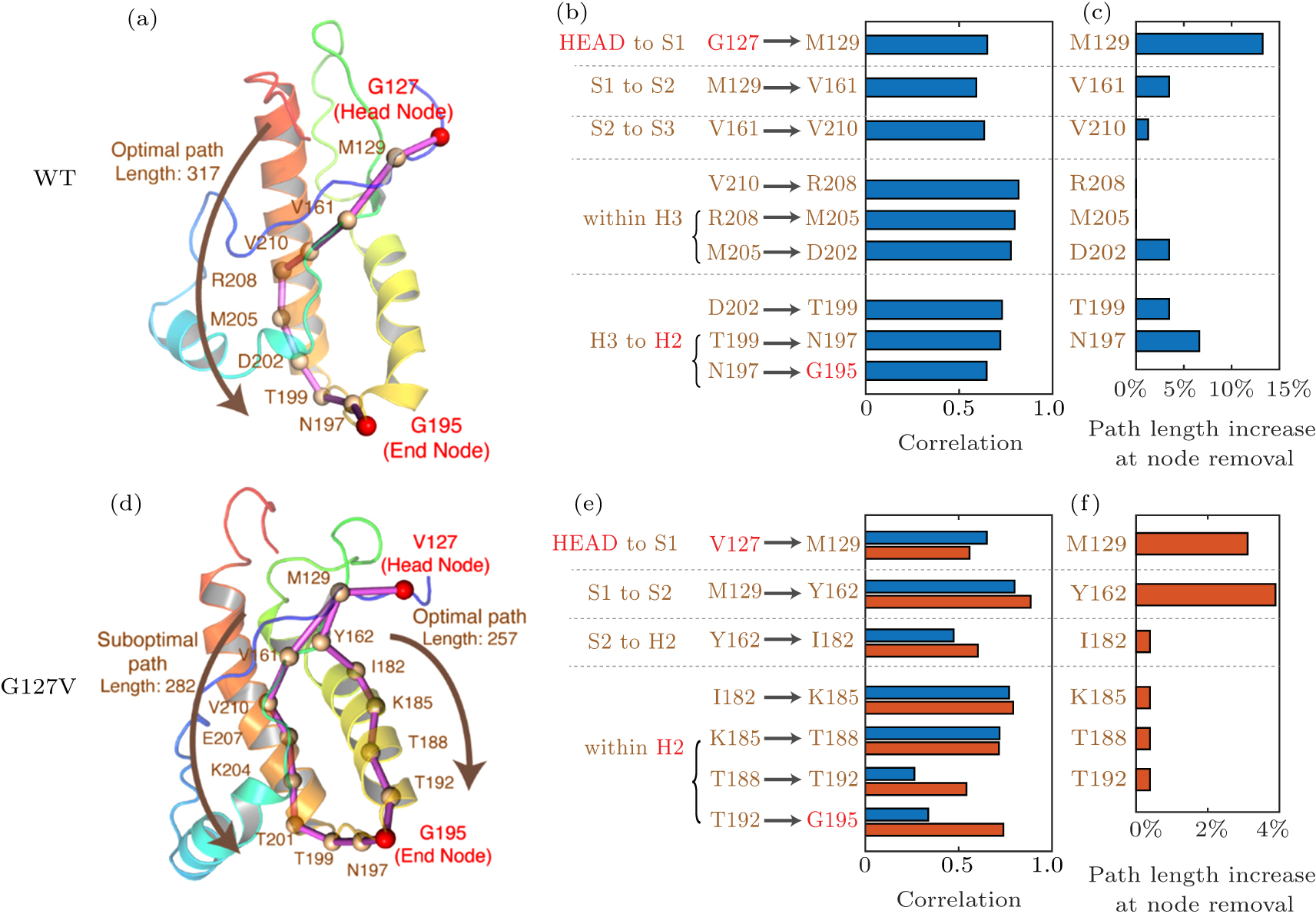
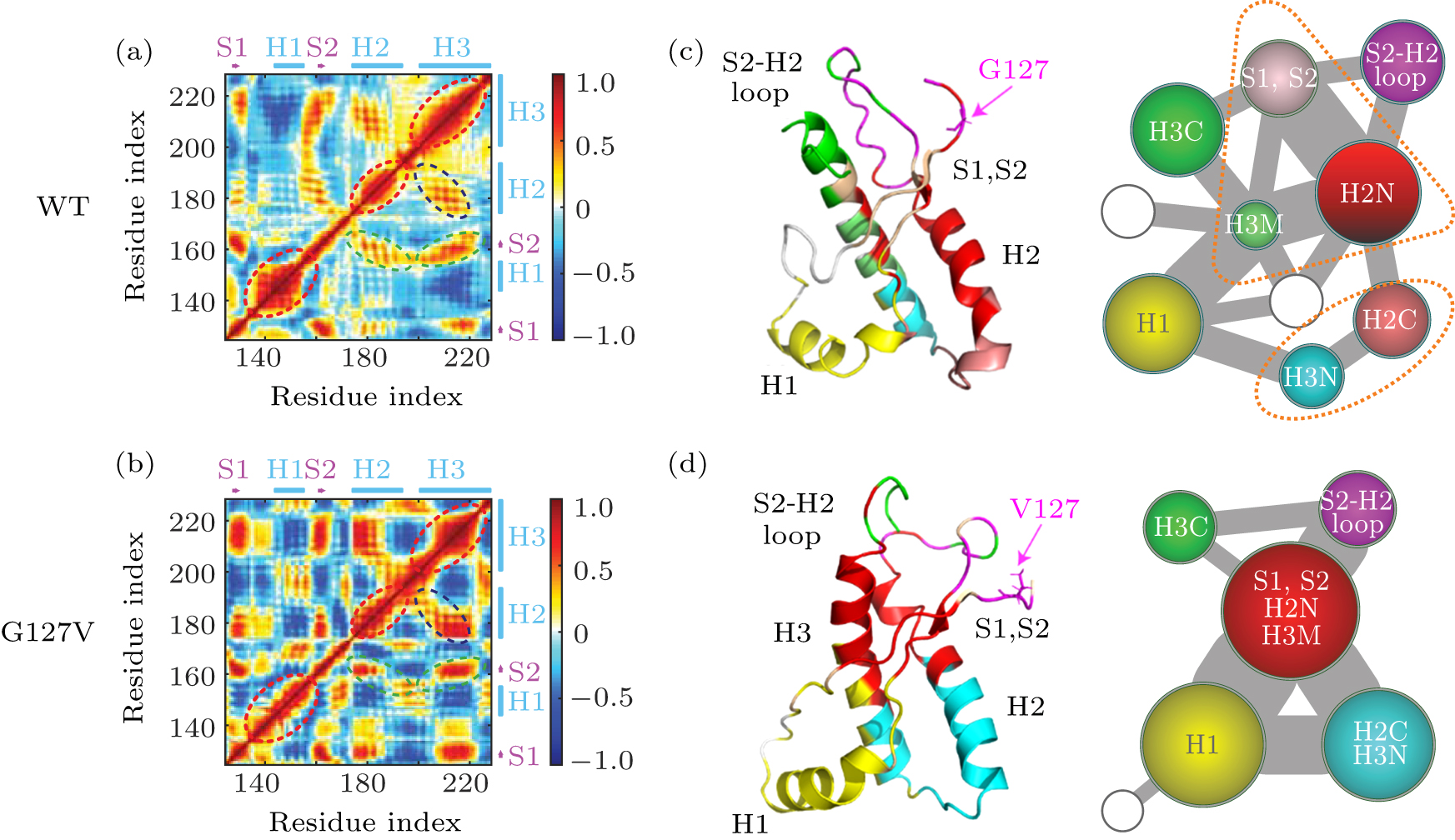
Abstract Prion diseases are associated with the misfolding of the normal helical cellular form of prion protein (PrPC) into the β-sheet-rich scrapie form (PrPSc) and the subsequent aggregation of PrPSc into amyloid fibrils. Recent studies demonstrated that a naturally occurring variant V127 of human PrPC is intrinsically resistant to prion conversion and aggregation, and can completely prevent prion diseases. However, the underlying molecular mechanism remains elusive. Herein we perform multiple microsecond molecular dynamics simulations on both wildtype (WT) and V127 variant of human PrPC to understand at atomic level the protective effect of V127 variant. Our simulations show that G127V mutation not only increases the rigidity of the S2–H2 loop between strand-2 (S2) and helix-2 (H2), but also allosterically enhances the stability of the H2 C-terminal region. Interestingly, previous studies reported that animals with rigid S2–H2 loop usually do not develop prion diseases, and the increase in H2 C-terminal stability can prevent misfolding and oligomerization of prion protein. The allosteric paths from G/V127 to H2 C-terminal region are identified using dynamical network analyses. Moreover, community network analyses illustrate that G127V mutation enhances the global correlations and intra-molecular interactions of PrP, thus stabilizing the overall PrPC structure and inhibiting its conversion into PrPSc. This study provides mechanistic understanding of human V127 variant in preventing prion conversion which may be helpful for the rational design of potent anti-prion compounds.
|
Received: 24 June 2020
Revised: 19 July 2020
Accepted manuscript online: 28 July 2020
|
|
PACS:
|
87.15.-v
|
(Biomolecules: structure and physical properties)
|
| |
87.14.E-
|
(Proteins)
|
| |
87.15.ap
|
(Molecular dynamics simulation)
|
|
|
Corresponding Authors:
†Corresponding author. E-mail: ghwei@fudan.edu.cn
|
| About author: †Corresponding author. E-mail: ghwei@fudan.edu.cn * Project supported by the Key Program of the National Key Research and Development Program of China (Grant No. 2016YFA0501702) and the National Natural Science Foundation of China (Grant No. 11674065). |
Cite this article:
Yiming Tang(唐一鸣), Yifei Yao(姚逸飞), and Guanghong Wei(韦广红)† Structural and dynamical mechanisms of a naturally occurring variant of the human prion protein in preventing prion conversion 2020 Chin. Phys. B 29 108710
|
| [1] |
|
| [2] |
|
| [3] |
|
| [4] |
|
| [5] |
Will R G, Ironside J W, Zeidler M, Cousens S N, Estibeiro K, Alperovitch A, Poser S, Pocchiari M, Hofmar A, Smith P G 1996 Lancet 347 921 DOI: 10.1016/S0140-6736(96)91412-9 |
| [6] |
|
| [7] |
Lugaresi E, Medori R, Montagna P, Baruzzi A, Cortelli P, Lugaresi A, Tinuper P, Zucconi M, Gambetti P 1986 N. Engl. J. Med. 315 997 DOI: 10.1056/NEJM198610163151605 |
| [8] |
Medori R, Tritschler H-J, LeBlanc A, Villare F, Manetto V, Chen H Y, Xue R, Leal S, Montagna P, Cortelli P, Tinuper P, Avoni P, Mochi M, Baruzzi A, Hauw J J, Ott J, Lugaresi E, Autilio-Gambetti L, Gambetti P 1992 N. Engl. J. Med. 326 444 DOI: 10.1056/NEJM199202133260704 |
| [9] |
|
| [10] |
Matthews J D, Glasse R, Lindenbaum S 1968 Lancet 2 449
|
| [11] |
|
| [12] |
Farquhar C F, Somerville R A, Bruce M E 1998 Nature 391 345 DOI: 10.1038/34818 |
| [13] |
|
| [14] |
|
| [15] |
|
| [16] |
Tuite M F, Serio T R 2010 Nat. Rev. Mol. Cell Biol. 11 823 DOI: 10.1038/nrm3007 |
| [17] |
|
| [18] |
Nussbaum J M, Schilling S, Cynis H, Silva A, Swanson E, Wangsanut T, Tayler K, Wiltgen B, Hatami A, Rönicke R, Reymann K, Hutter-Paier B, Alexandru A, Jagla W, Graubner S, Glabe C G, Demuth H U, Bloom G S 2012 Nature 485 651 DOI: 10.1038/nature11060 |
| [19] |
|
| [20] |
Masuda-Suzukake M, Nonaka T, Hosokawa M, Oikawa T, Arai T, Akiyama H, Mann D M A, Hasegawa M 2013 Brain 136 1128 DOI: 10.1093/brain/awt037 |
| [21] |
|
| [22] |
|
| [23] |
|
| [24] |
|
| [25] |
|
| [26] |
|
| [27] |
|
| [28] |
Zahn R, Liu A, Lührs T, Riek R, Von Schroetter C, Garcia F L, Billeter M, Calzolai L, Wider G, Wüthrich K 2000 Proc. Natl. Acad. Sci. USA 97 145 DOI: 10.1073/pnas.97.1.145 |
| [29] |
Calzolai L, Lysek D A, Güntert P, Von Schroetter C, Riek R, Zahn R, Wüthrich K 2000 Proc. Natl. Acad. Sci. USA 97 8340 DOI: 10.1073/pnas.97.15.8340 |
| [30] |
Knaus K J, Morillas M, Swietnicki W, Malone M, Surewicz W K, Yee V C 2001 Nat. Struct. Biol. 8 770 DOI: 10.1038/nsb0901-770 |
| [31] |
|
| [32] |
Provenzano L, Ryan Y, Hilton D A, Lyons-Rimmer J, Dave F, Maze E A, Adams C L, Rigby-Jones R, Ammoun S, Hanemann C O 2017 Oncogene 36 6132 DOI: 10.1038/onc.2017.200 |
| [33] |
Linsenmeier L, Altmeppen H C, Wetzel S, Mohammadi B, Saftig P, Glatzel M 2017 Biochim. Biophys. Acta-Mol. Cel. Res. 1864 2128 DOI: 10.1016/j.bbamcr.2017.06.022 |
| [34] |
|
| [35] |
|
| [36] |
Franzmann T M, Jahnel M, Pozniakovsky A, Mahamid J, Holehouse A S, Nüske E, Richter D, Baumeister W, Grill S W, Pappu R V, Hyman A A, Alberti S 2018 Science 359 6371 DOI: 10.1126/science.aao5654 |
| [37] |
Pan K M, Baldwin M, Nguyen J, Gasset M, Serban A, Groth D, Mehlhorn I, Huang Z, Fletterick R J, Cohen F E, Prusiner S B 1993 Proc. Natl. Acad. Sci. USA 90 10962 DOI: 10.1073/pnas.90.23.10962 |
| [38] |
|
| [39] |
|
| [40] |
|
| [41] |
|
| [42] |
|
| [43] |
|
| [44] |
Petersen R B, Parchi P, Richardson S L, Urig C B, Gambetti P 1996 J. Biol. Chem. 271 12661 DOI: 10.1074/jbc.271.21.12661 |
| [45] |
Zarranz J J, Digon A, Atarés B, Rodríguez-Martinez A B, Arce A, Carrera N, Fernández-Manchola I, Fernández-Martínez M, Fernández-Maiztegui C, Forcadas I, Galdos L, Gómez-Esteban J C, Ibáñez A, Lezcano E, De López Munain A, Martí-Massó J F, Mendibe M M, Urtasun M, Uterga J M, Saracibar N, Velasco F, De Pancorbo M M 2005 J. Neurol. Neurosurg. Psychiatry 76 1491 DOI: 10.1136/jnnp.2004.056606 |
| [46] |
Woulfe J, Kertesz A, Frohn I, Bauer S, George-Hyslop P S, Bergeron C 2005 Acta Neuropathol. 110 317 DOI: 10.1007/s00401-005-1054-0 |
| [47] |
Tartaglia M C, Thai J N, See T, Kuo A, Harbaugh R, Raudabaugh B, Cali I, Sattavat M, Sanchez H, DeArmond S J, Geschwind M D 2010 J. Neuropathol. Exp. Neurol. 69 1220 DOI: 10.1097/NEN.0b013e3181ffc39c |
| [48] |
Palmer M S, Dryden A J, Hughes J T, Collinge J 1991 Nature 352 340 DOI: 10.1038/352340a0 |
| [49] |
Shibuya S, Higuchi J, Shin R W, Tateishi J, Kitamoto T 1998 Ann. Neurol. 43 826 DOI: 10.1002/ana.v43:6 |
| [50] |
Soldevila M, Calafell F, Andrés A M, Yagüe J, Helgason A, Stefánsson K, Bertranpetit J 2003 Hum. Mutat. 22 104 DOI: 10.1126/science.aao5654 |
| [51] |
|
| [52] |
Mead S, Whitfield J, Poulter M, Shah P, Uphill J, Campbell T, Al-Dujaily H, Hummerich H, Beck J, Mein C A, Verzilli C, Whittaker J, Alpers M P, Collinge J 2009 N. Engl. J. Med. 361 2056 DOI: 10.1056/NEJMoa0809716 |
| [53] |
Asante E A, Smidak M, Grimshaw A, Houghton R, Tomlinson A, Jeelani A, Jakubcova T, Hamdan S, Richard-Londt A, Linehan J M, Brandner S, Alpers M, Whitfield J, Mead S, Wadsworth J D F, Collinge J 2015 Nature 522 478 DOI: 10.1038/nature14510 |
| [54] |
|
| [55] |
Pan A C, Jacobson D, Yatsenko K, Sritharan D, Weinreich T M, Shaw D E 2019 Proc. Natl. Acad. Sci. USA 116 4244 DOI: 10.1073/pnas.1815431116 |
| [56] |
|
| [57] |
|
| [58] |
|
| [59] |
|
| [60] |
|
| [61] |
|
| [62] |
Mandujano-Rosas L A, Osorio-González D, Reyes-Romero P G, Mulia-Rodríguez J 2014 Open J. Biophys. 04 169 DOI: 10.4236/ojbiphy.2014.44016 |
| [63] |
|
| [64] |
|
| [65] |
Barducci A, Chelli R, Procacci P, Schettino V, Gervasio F L, Parrinello M 2006 J. Am. Chem. Soc. 128 2705 DOI: 10.1021/ja057076l |
| [66] |
Caldarulo E, Barducci A, Wüthrich K, Parrinello M 2017 Proc. Natl. Acad. Sci. USA 114 9617 DOI: 10.1073/pnas.1712155114 |
| [67] |
|
| [68] |
|
| [69] |
Kurt T D, Aguilar-Calvo P, Jiang L, Rodriguez J A, Alderson N, Eisenberg D S, Sigurdson C J 2017 J. Biol. Chem. 292 19076 DOI: 10.1074/jbc.M117.794107 |
| [70] |
Sigurdson C J, Nilsson K P R, Hornemann S, Manco G, Fernández-Borges N, Schwarz P, Castilla J, Wüthrich K, Aguzzi A 2010 J. Clin. Invest. 120 2590 DOI: 10.1172/JCI42051 |
| [71] |
|
| [72] |
Gossert A D, Bonjour S, Lysek D A, Fiorito F, Wüthrich K 2005 Proc. Natl. Acad. Sci. USA 102 646 DOI: 10.1073/pnas.0409008102 |
| [73] |
|
| [74] |
|
| [75] |
Agarwal S, Döring K, Gierusz L A, Iyer P, Lane F M, Graham J F, Goldmann W, Pinheiro T J T, Gill A C 2015 Sci. Rep. 5 15528 DOI: 10.1038/srep15528 |
| [76] |
Kaneko K, Zulianello L, Scott M, Cooper C M, Wallace A C, James T L, Cohen F E, Prusiner S B 1997 Proc. Natl. Acad. Sci. USA 94 10069 DOI: 10.1073/pnas.94.19.10069 |
| [77] |
Knaus K J, Morillas M, Swietnicki W, Malone M, Surewicz W K, Yee V C 2001 Nat. Struct. Biol. 8 770 DOI: 10.1038/nsb0901-770 |
| [78] |
Bjorndahl T C, Zhou G P, Liu X, Perez-Pineiro R, Semenchenko V, Saleem F, Acharya S, Bujold A, Sobsey C A, Wishart D S 2011 Biochemistry 50 1162 DOI: 10.1021/bi101435c |
| [79] |
Singh J, Kumar H, Sabareesan A T, Udgaonkar J B 2014 J. Am. Chem. Soc. 136 16704 DOI: 10.1021/ja510964t |
| [80] |
|
| [81] |
Huang J J, Li X N, Liu W L, Yuan H Y, Gao Y, Wang K, Tang B, Pang D W, Chen J, Liang Y 2020 J. Mol. Biol. 432 828 DOI: 10.1016/j.jmb.2019.11.020 |
| [82] |
|
| [83] |
|
| [84] |
Miao Y, Nichols S E, Gasper P M, Metzger V T, McCammon J A 2013 Proc. Natl. Acad. Sci. USA 110 10982 DOI: 10.1073/pnas.1309755110 |
| [85] |
|
| [86] |
|
| [87] |
|
| [88] |
|
| [89] |
|
| [90] |
|
| [91] |
|
| [92] |
Alcalá-Corona S A, Velázquez-Caldelas T E, Espinal-Enríquez J, Hernández-Lemus E 2016 Front. Physiol. 7 184 DOI: 10.3389/fphys.2016.00184 |
| [93] |
Guo C, Zhou H X 2016 Proc. Natl. Acad. Sci. USA 113 E6776
|
| [94] |
|
| [95] |
The PyMOL Molecular Graphics System, Version 2.0 Schrödinger, LLC
|
| [96] |
Lindorff-Larsen K, Piana S, Palmo K, Maragakis P, Klepeis J L, Dror R O, Shaw D E 2010 Proteins Struct. Funct. Bioinforma. 78 1950 DOI: 10.1002/prot.22711 |
| [97] |
|
| [98] |
|
| [99] |
Bussi G, Donadio D, Parrinello M 2007 J. Chem. Phys. 126 014101 DOI: 10.1063/1.2408420 |
| [100] |
|
| [101] |
|
| [102] |
|
| [103] |
|
| [104] |
|
| No Suggested Reading articles found! |
|
|
Viewed |
|
|
|
Full text
|
|
|
|
|
Abstract
|
|
|
|
|
Cited |
|
|
|
|
Altmetric
|
|
blogs
Facebook pages
Wikipedia page
Google+ users
|
Online attention
Altmetric calculates a score based on the online attention an article receives. Each coloured thread in the circle represents a different type of online attention. The number in the centre is the Altmetric score. Social media and mainstream news media are the main sources that calculate the score. Reference managers such as Mendeley are also tracked but do not contribute to the score. Older articles often score higher because they have had more time to get noticed. To account for this, Altmetric has included the context data for other articles of a similar age.
View more on Altmetrics
|
|
|