Please wait a minute...
Chin. Phys. B, 2020, Vol. 29(10): 108709    DOI: 10.1088/1674-1056/abaee1
Special Issue: SPECIAL TOPIC — Modeling and simulations for the structures and functions of proteins and nucleic acids
TOPICAL REVIEW—Modeling and simulations for the structures and functions of proteins and nucleic acids Prev   Next  

Twisting mode of supercoil leucine-rich domain mediates peptide sensing in FLS2–flg22–BAK1 complex

Zhi-Chao Liu(刘志超)1,3, Qin Liu(刘琴)4, Chan-You Chen(陈禅友)4, Chen Zeng(曾辰)3, Peng Ran(冉鹏)1,3, Yun-Jie Zhao(赵蕴杰)2,†, and Lei Pan(潘磊)4,
1 School of Biological Information, Chongqing University of Posts and Telecommunications, Chongqing 400065, China
2 Institute of Biophysics and Department of Physics, Central China Normal University, Wuhan 430079, China
3 Department of Physics, The George Washington University, Washington, DC, 20052, USA
4 School of Life Sciences, Jianghan University, Wuhan 430056, China
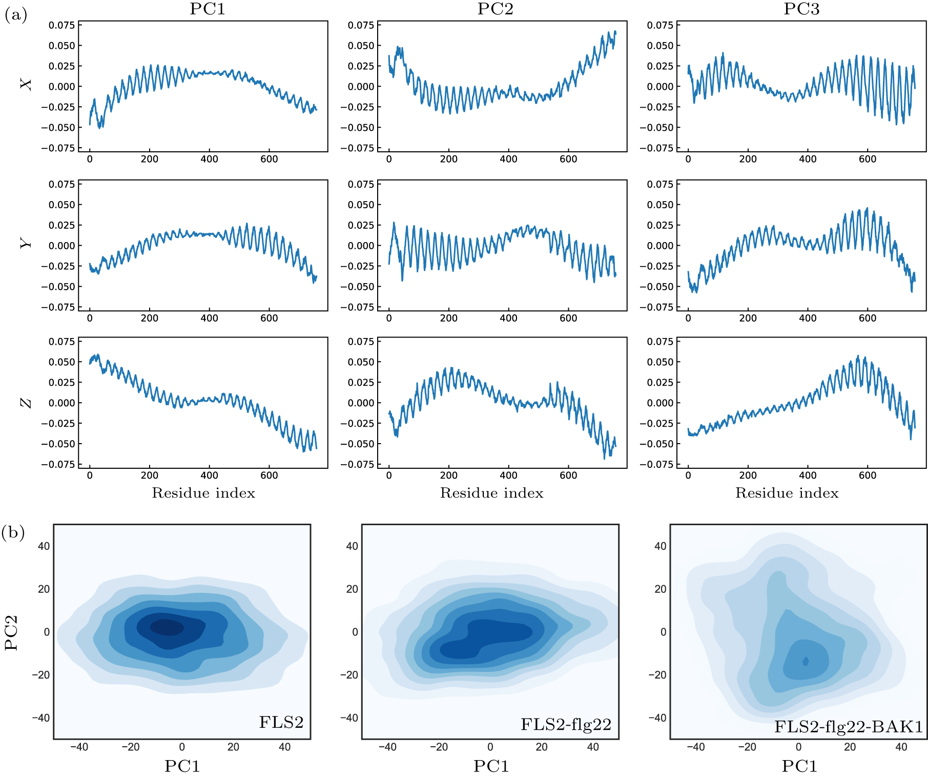
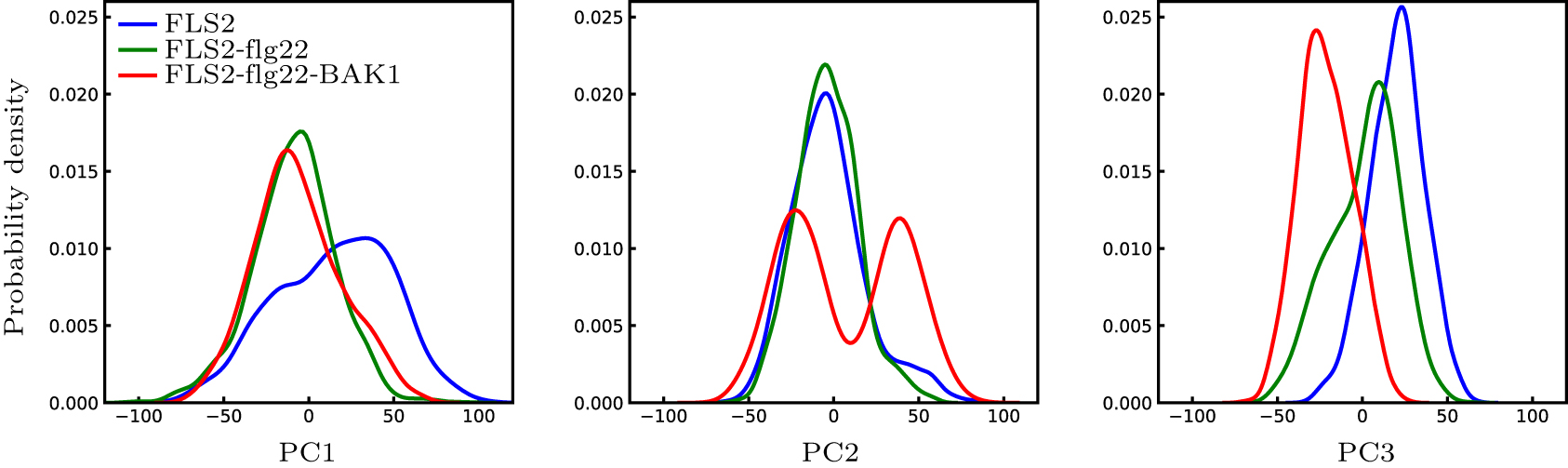
Abstract  

Plants and animals recognize microbial invaders by detecting pathogen-associated molecular patterns (PAMPs) through pattern-recognition receptors (PRRs). This recognition plays a crucial role in plant immunity. The newly discovered protein in plants that responds to bacterial flagellin, i.e., flagellin-sensitive 2 (FLS2), is ubiquitously expressed and present in many plants. The association of FLS2 and BAK1, facilitated by a highly conserved epitope flg22 of flagellin, triggers such downstream immune responses as activated MAPK pathway and elevated reactive oxygen species (ROS) for bacterial defense and plant immunity. Here we study the intrinsic dynamics and conformational change of FLS2 upon the formation of the FLS2–flg22–BAK1 complex. The top intrinsic normal modes and principal structural fluctuation components are very similar, showing two bending modes and one twisting mode. The twisting mode alone, however, accounts for most of the conformational change of FLS2 induced by binding with flg22 and BAK1. This study indicates that flg22 binding suppresses FLS2 conformational fluctuation, especially on the twisting motion, thus facilitating FLS2–BAK1 interaction. A detailed analysis of this sensing mechanism may aid better design on both PRR and peptide mimetics for plant immunity.

Keywords:  plant immunity      flagellin-sensitive 2      peptide sensing mechanism  
Received:  23 June 2020      Revised:  07 August 2020      Accepted manuscript online:  13 August 2020
PACS:  87.14.gn (RNA)  
  87.15.K- (Molecular interactions; membrane-protein interactions)  
  87.10.Ca (Analytical theories)  
  87.15.A- (Theory, modeling, and computer simulation)  
Corresponding Authors:  Corresponding author. E-mail: yjzhaowh@mail.ccnu.edu.cn Corresponding author. E-mail: leipan@jhun.edu.cn   
About author: 
†Corresponding author. E-mail: yjzhaowh@mail.ccnu.edu.cn
‡Corresponding author. E-mail: leipan@jhun.edu.cn
* Project supported by the National Natural Science Foundation of China (Grant No. 11704140), self-determined research funds of CCNU from the Colleges’ Basic Research and Operation of MOE (Grant No. CCNU20TS004) (Y. Z.), and the China Scholarship Council Fund (Grant No. 201708420039) (L. P.).

Cite this article: 

Zhi-Chao Liu(刘志超), Qin Liu(刘琴), Chan-You Chen(陈禅友), Chen Zeng(曾辰), Peng Ran(冉鹏), Yun-Jie Zhao(赵蕴杰)†, and Lei Pan(潘磊)‡ Twisting mode of supercoil leucine-rich domain mediates peptide sensing in FLS2–flg22–BAK1 complex 2020 Chin. Phys. B 29 108709

Fig. 1.  

FLS2–BAK1 interface correlation is enhanced upon the flg22 binding. The horizontal axis indicates the interface residues in BAK1, and the vertical axis shows the interface residues in FLS2. (a) Interface correlation of FLS2–BAK1 without flg22; (b) with flg22.

Fig. 2.  

(a), (c) Pitch length and (b), (d) radius distribution of FLS2 superhelix structure for FLS2, FLS2–flg22, and FLS2–flg22–BAK1 at the start (upper panel) and end (lower panel) episodes of the whole 200 ns MD simulations. From left to right, pitch length and its variance decrease. The radius also fluctuates less and slightly decreases.

Fig. 3.  

First 3 dominant normal modes of FLS2. (a) Left: mode 1 (side view) shows the bending motion with two ends moving asynchronously; Middle: mode 2 (top view) shows the twisting motion with two ends rotating around the center axis in opposite directions; Right: mode 3 (side view) shows another bending motion with two synchronous ends. (b) The x, y, z components (in normalized scale) of the first three normal modes (NMs).

Fig. 4.  

Principal component analysis of FLS2 MD trajectory. (a) The x, y, z dimensions of the first three principal components. PC1 and PC2 indicate bending modes and PC3 twisting mode of helix. (b) The FLS2 structure distribution density of the MD trajectories in the 2D space spanned by PC1 and PC2 for FLS2-only (lower left), FLS2–flg22 (lower middle), and FLS2–flg22–BAK1 (lower right).

Fig. 5.  

(a) Explained variance ratio by the top 10 PCs in PCA. Top 3 components (bending1, bending2, and twisting mode) contribute to around 70% of the variance; (b) Correlation of the top 3 components of PCA and the top 3 modes of ANM. PC1 matches best with NM1 (Pearson correlation coefficient 0.96); PC2 corresponds to NM3 (0.93); PC3 to NM2 (0.86). (c) Top PCs and NMs projection correlation. The projection coefficients of the FLS2 MD structures correlate well for PC1–NM1 (left), PC2–NM3 (center), and PC3–NM2 (right), with the correlation coefficients indicated.

Fig. 6.  

Top PCs and NMs projection distribution. The distribution of projection coefficients of FLS2 structure in (a) PC1, (b) PC2, and (c) PC3 direction. The FLS2 structure in the three different complexes shows no obvious difference in PC1 and PC2 directions while shows an obvious global shift for FLS2, FLS2–flg22, and FLS2–flg22–BAK1 conditions, with FLS2–flg22 being the intermediate state.

Fig. 7.  

Proposed interaction mechanism of the FLS2–flg22–BAK1 complex. (a) Interaction of FLS2–flg22–BAK1. N- and C-terminal ends of flg22 interact with At-FLS2 and promote the binding of BAK1. Interaction of FLS2–flg15–BAK1 in (b) Arabidopsis thaliana (At) and (c) Solanum lycopersicum (Sl). The new N-terminus of flg15 fails to bind with At-FLS2, thus resulting in little restriction on the twisting dynamics of FLS2 and, therefore much weaker FLS2–BAK1 association or correspondingly, much weaker downstream immune response. In Sl, the N-terminus of flg15 binds with Sl-FLS2 to induce a tight binding of BAK1.

Fig. 8.  

Conservation analysis of FLS2–flg22 binding sites. (a) Conservation of flg22. The N-terminus (first 7 residues) is less conserved than the C-terminus. (b) The corresponding binding sites of FLS2 with flg22 N-terminus are also less conserved than those with flg22 C-terminus.

[1]
Helft L, Thompson M, Bent A F 2016 PLoS One 11 e0157155 DOI: 10.1371/journal.pone.0157155
[2]
Saijo Y, Loo E P, Yasuda S 2018 Plant J. 93 592 DOI: 10.1111/tpj.2018.93.issue-4
[3]
Medzhitov R, Janeway C A Jr. 1997 Cell 91 295 DOI: 10.1016/S0092-8674(00)80412-2
[4]
Boller T, Felix G 2009 Annu. Rev. Plant Biol. 60 379 DOI: 10.1146/annurev.arplant.57.032905.105346
[5]
Zipfel C 2014 Trends Immunol 35 345 DOI: 10.1016/j.it.2014.05.004
[6]
Boutrot F, Zipfel C 2017 Annu Rev. Phytopathol 55 257 DOI: 10.1146/annurev-phyto-080614-120106
[7]
Zipfel C 2009 Curr. Opin. Plant. Biol. 12 414 DOI: 10.1016/j.pbi.2009.06.003
[8]
Tintor N, Ross A, Kanehara K, Yamada K, Fan L, Kemmerling B, Nurnberger T, Tsuda K, Saijo Y 2013 Proc. Natl. Acad. Sci. USA 110 6211 DOI: 10.1073/pnas.1216780110
[9]
Robatzek S, Wirthmueller L 2013 Protoplasma 250 671 DOI: 10.1007/s00709-012-0459-6
[10]
Sun Y, Li L, Macho A P, Han Z, Hu Z, Zipfel C, Zhou J M, Chai J 2013 Science 342 624 DOI: 10.1126/science.1243825
[11]
Padmanabhan M, Cournoyer P, Dinesh-Kumar S P 2009 Cell Microbiol 11 191 DOI: 10.1111/cmi.2009.11.issue-2
[12]
Zhang Z, Thomma B P 2013 J. Integr. Plant Biol. 55 1212 DOI: 10.1111/jipb.12080
[13]
Chinchilla D, Bauer Z, Regenass M, Boller T, Felix G 2006 Plant Cell 18 465 DOI: 10.1105/tpc.105.036574
[14]
Gomez-Gomez L, Boller T 2000 Mol. Cell 5 1003 DOI: 10.1016/S1097-2765(00)80265-8
[15]
Zipfel C, Kunze G, Chinchilla D, Caniard A, Jones J D, Boller T, Felix G 2006 Cell 125 749 DOI: 10.1016/j.cell.2006.03.037
[16]
Mithoe S C, Menke F L 2018 Curr. Opin. Plant Biol. 45 162 DOI: 10.1016/j.pbi.2018.07.008
[17]
Chinchilla D, Zipfel C, Robatzek S, Kemmerling B, Nurnberger T, Jones J D, Felix G, Boller T 2007 Nature 448 497 DOI: 10.1038/nature05999
[18]
Somssich M, Ma Q, Weidtkamp-Peters S, Stahl Y, Felekyan S, Bleckmann A, Seidel C A, Simon R 2015 Sci. Signal 8 ra76 DOI: 10.1126/scisignal.aab0598
[19]
Beck M, Zhou J, Faulkner C, MacLean D, Robatzek S 2012 Plant Cell 24 4205 DOI: 10.1105/tpc.112.100263
[20]
Fischer E 1894 Berichte der deutschen chemischen Gesellschaft 27 2985 DOI: 10.1002/(ISSN)1099-0682
[21]
Koshland D E 1958 Proc. Natl. Acad. Sci. USA 44 98 DOI: 10.1073/pnas.44.2.98
[22]
Tobi D, Bahar I 2005 Proc. Natl. Acad. Sci. USA 102 18908 DOI: 10.1073/pnas.0507603102
[23]
Bakan A, Bahar I 2009 Proc. Natl. Acad. Sci. USA 106 14349 DOI: 10.1073/pnas.0904214106
[24]
Ponzoni L, Bahar I 2018 Proc. Natl. Acad. Sci. USA 115 4164 DOI: 10.1073/pnas.1715896115
[25]
Wang K L, Jian Y R, Wang H W, Zeng C, Zhao Y J 2018 Bioinformatics 34 3131 DOI: 10.1093/bioinformatics/bty345
[26]
Wang H W, Wang K L, Guan Z Y, Jian Y R, Jia Y, Kashanchi F, Zeng C, Zhao Y J 2017 Chin. Phys. B 26 128702 DOI: 10.1088/1674-1056/26/12/128702
[27]
Zhao Y, Jian Y, Liu Z, Liu H, Liu Q, Chen C, Li Z, Wang L, Huang H H, Zeng C 2017 Sci. Rep. 7 2876 DOI: 10.1038/s41598-017-03003-5
[28]
Chen H, Zhao Y J, Li H T, Zhang D Y, Huang Y Z, Shen Q, Van Duyne R, Kashanchi F, Zeng C, Liu S Y 2014 Plos One 9 DOI: 10.1371/journal.pone.0109154
[29]
Yi Yang Q G, Zhang B G, Shi Y Z, Shao Z G 2018 Chin. Phys. B 27 38701 DOI: 10.1088/1674-1056/27/3/038701
[30]
Dill K A, Chan H S 1997 Nat. Struct. Biol. 4 10 DOI: 10.1038/nsb0197-10
[31]
Brunette T J, Parmeggiani F, Huang P S, Bhabha G, Ekiert D C, Tsutakawa S E, Hura G L, Tainer J A, Baker D 2015 Nature 528 580 DOI: 10.1038/nature16162
[32]
Enkhbayar P, Kamiya M, Osaki M, Matsumoto T, Matsushima N 2004 Proteins 54 394 DOI: 10.1002/prot.10605
[33]
Stumpp M T, Forrer P, Binz H K, Pluckthun A 2003 J. Mol. Biol. 332 471 DOI: 10.1016/S0022-2836(03)00897-0
[34]
Ramisch S, Weininger U, Martinsson J, Akke M, Andre I 2014 Proc. Natl. Acad Sci. USA 111 17875 DOI: 10.1073/pnas.1413638111
[35]
Doyle L, Hallinan J, Bolduc J, Parmeggiani F, Baker D, Stoddard B L, Bradley P 2015 Nature 528 585 DOI: 10.1038/nature16191
[36]
Emberly E G, Mukhopadhyay R, Wingreen N S, Tang C 2003 J. Mol. Biol. 327 229 DOI: 10.1016/S0022-2836(03)00097-4
[37]
Van Der Spoel D, Lindahl E, Hess B, Groenhof G, Mark A E, Berendsen H J 2005 J. Comput. Chem. 26 1701 DOI: 10.1002/(ISSN)1096-987X
[38]
Duan Y, Wu C, Chowdhury S, Lee M C, Xiong G, Zhang W, Yang R, Cieplak P, Luo R, Lee T, Caldwell J, Wang J, Kollman P 2003 J. Comput. Chem. 24 1999 DOI: 10.1002/(ISSN)1096-987X
[39]
Sethi A, Eargle J, Black A A, Luthey-Schulten Z 2009 Proc. Natl. Acad. Sci. USA 106 6620 DOI: 10.1073/pnas.0810961106
[40]
Humphrey W, Dalke A, Schulten K 1996 J. Mol. Graph 14 33 DOI: 10.1016/0263-7855(96)00018-5
[41]
Enkhbayar P, Damdinsuren S, Osaki M, Matsushima N 2008 Comput. Biol. Chem. 32 307 DOI: 10.1016/j.compbiolchem.2008.03.012
[42]
Atilgan A R, Durell S R, Jernigan R L, Demirel M C, Keskin O, Bahar I 2001 Biophys. J. 80 505 DOI: 10.1016/S0006-3495(01)76033-X
[43]
Eyal E, Lum G, Bahar I 2015 Bioinformatics 31 1487 DOI: 10.1093/bioinformatics/btu847
[44]
Ashkenazy H, Erez E, Martz E, Pupko T, Ben-Tal N 2010 Nucleic Acids Res. 38 W529 DOI: 10.1093/nar/gkq399
[45]
Tang Q Y, Zhang Y Y, Wang J, Wang W, Chialvo D R 2017 Phys. Rev. Lett. 118 088102 DOI: 10.1103/PhysRevLett.118.088102
[46]
Tang Q Y, Kaneko K 2020 PLoS Comput. Biol. 16 e1007670 DOI: 10.1371/journal.pcbi.1007670
[47]
Bahar I, Lezon T R, Yang L W, Eyal E 2010 Ann. Rev. Biophys. 39 23 DOI: 10.1146/annurev.biophys.093008.131258
[48]
Mueller K, Bittel P, Chinchilla D, Jehle A K, Albert M, Boller T, Felix G 2012 Plant Cell 24 2213 DOI: 10.1105/tpc.112.096073
[49]
Dunning F M, Sun W, Jansen K L, Helft L, Bent A F 2007 Plant Cell 19 3297 DOI: 10.1105/tpc.106.048801
[1] RNAGCN: RNA tertiary structure assessment with a graph convolutional network
Chengwei Deng(邓成伟), Yunxin Tang(唐蕴芯), Jian Zhang(张建), Wenfei Li(李文飞), Jun Wang(王骏), and Wei Wang(王炜). Chin. Phys. B, 2022, 31(11): 118702.
[2] Force-constant-decayed anisotropic network model: An improved method for predicting RNA flexibility
Wei-Bu Wang(王韦卜), Xing-Yuan Li(李兴元), and Ji-Guo Su(苏计国). Chin. Phys. B, 2022, 31(6): 068704.
[3] Single-molecule mechanical folding and unfolding kinetics of armless mitochondrial tRNAArg from Romanomermis culicivorax
Yan-Hui Li(李彦慧), Zhen-Sheng Zhong(钟振声), and Jie Ma(马杰). Chin. Phys. B, 2021, 30(10): 108203.
[4] Enhancements of the Gaussian network model in describing nucleotide residue fluctuations for RNA
Wen-Jing Wang(王文静) and Ji-Guo Su(苏计国). Chin. Phys. B, 2021, 30(5): 058701.
[5] Statistical potentials for 3D structure evaluation: From proteins to RNAs
Ya-Lan Tan(谭雅岚), Chen-Jie Feng(封晨洁), Xunxun Wang(王勋勋), Wenbing Zhang(张文炳), and Zhi-Jie Tan(谭志杰). Chin. Phys. B, 2021, 30(2): 028705.
[6] Computational prediction of RNA tertiary structures using machine learning methods
Bin Huang(黄斌), Yuanyang Du(杜渊洋), Shuai Zhang(张帅), Wenfei Li(李文飞), Jun Wang (王骏), and Jian Zhang(张建)†. Chin. Phys. B, 2020, 29(10): 108704.
[7] The theory of helix-based RNA folding kinetics and its application
Sha Gong(龚沙), Taigang Liu(刘太刚), Yanli Wang(王晏莉), and Wenbing Zhang(张文炳)†. Chin. Phys. B, 2020, 29(10): 108703.
[8] Methods and applications of RNA contact prediction
Huiwen Wang(王慧雯) and Yunjie Zhao(赵蕴杰)†. Chin. Phys. B, 2020, 29(10): 108708.
[9] Improving RNA secondary structure prediction using direct coupling analysis
Xiaoling He(何小玲), Jun Wang(王军), Jian Wang(王剑), Yi Xiao(肖奕). Chin. Phys. B, 2020, 29(7): 078702.
[10] A novel knowledge-based potential for RNA 3D structure evaluation
Yi Yang(杨毅), Qi Gu(辜琦), Ben-Gong Zhang(张本龚), Ya-Zhou Shi(时亚洲), Zhi-Gang Shao(邵志刚). Chin. Phys. B, 2018, 27(3): 038701.
[11] To what extent of ion neutralization can multivalent ion distributions around RNA-like macroions be described by Poisson-Boltzmann theory?
Gui Xiong(熊贵), Kun Xi(席昆), Xi Zhang(张曦), Zhi-Jie Tan(谭志杰). Chin. Phys. B, 2018, 27(1): 018203.
[12] Molecular dynamic simulation of the thermodynamic and kinetic properties of nucleotide base pair
Yu-Jie Wang(王宇杰), Zhen Wang(王珍), Yan-Li Wang(王晏莉), Wen-Bing Zhang(张文炳). Chin. Phys. B, 2017, 26(12): 128705.
[13] Theoretical studies on sRNA-mediated regulation in bacteria
Chang Xiao-Xue (常晓雪), Xu Liu-Fang (徐留芳), Shi Hua-Lin (史华林). Chin. Phys. B, 2015, 24(12): 128703.
[14] RNA structure prediction:Progress and perspective
Shi Ya-Zhou (时亚洲), Wu Yuan-Yan (吴园燕), Wang Feng-Hua (王凤华), Tan Zhi-Jie (谭志杰). Chin. Phys. B, 2014, 23(7): 078701.
No Suggested Reading articles found!