Please wait a minute...
Chin. Phys. B, 2020, Vol. 29(11): 110701    DOI: 10.1088/1674-1056/aba601
GENERAL Prev   Next  

Sensitivity enhancement of WS2-coated SPR-based optical fiber biosensor for detecting glucose concentration

Yun Cai(蔡云)1,4, Wei Li(李卫)1,2,3,4, †, Ye Feng(冯烨)1,4, Jian-Sheng Zhao(赵建胜)1,4, Gang Bai(白刚)1,4, Jie Xu(许杰)1,4, and Jin-Ze Li(李金泽)1,4$
1 College of Electronic and Optical Engineering & College of Microelectronics, Nanjing University of Posts and Telecommunications, Nanjing 210023, China
2 State Key Laboratory of Millimeter Waves, Southeast University, Nanjing 210093, China
3 State Key Laboratory of Luminescent Materials and Devices, South China University of Technology, Guangzhou 510641, China
4 Research Center of Optical Communications Engineering & Technology, Nanjing 210023, China
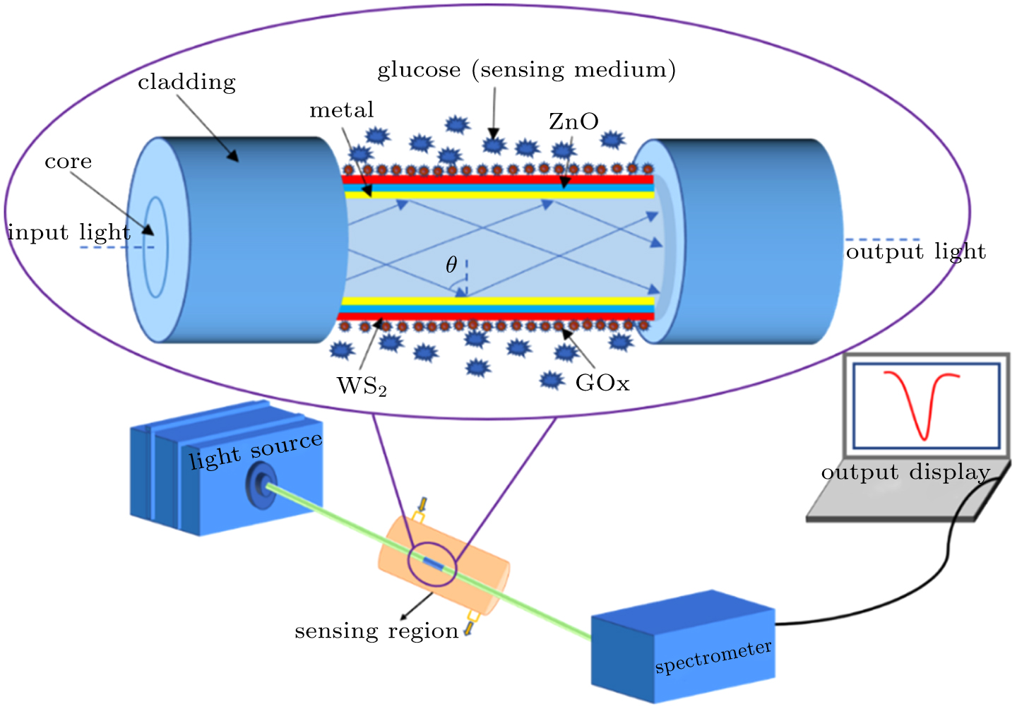
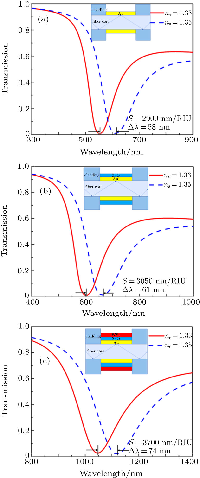
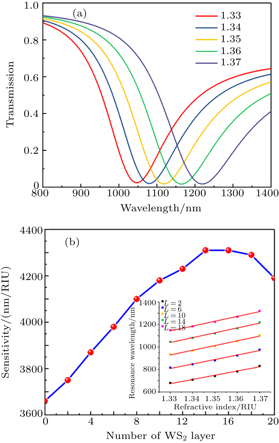
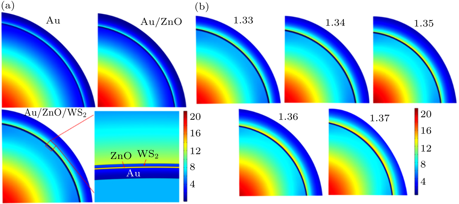
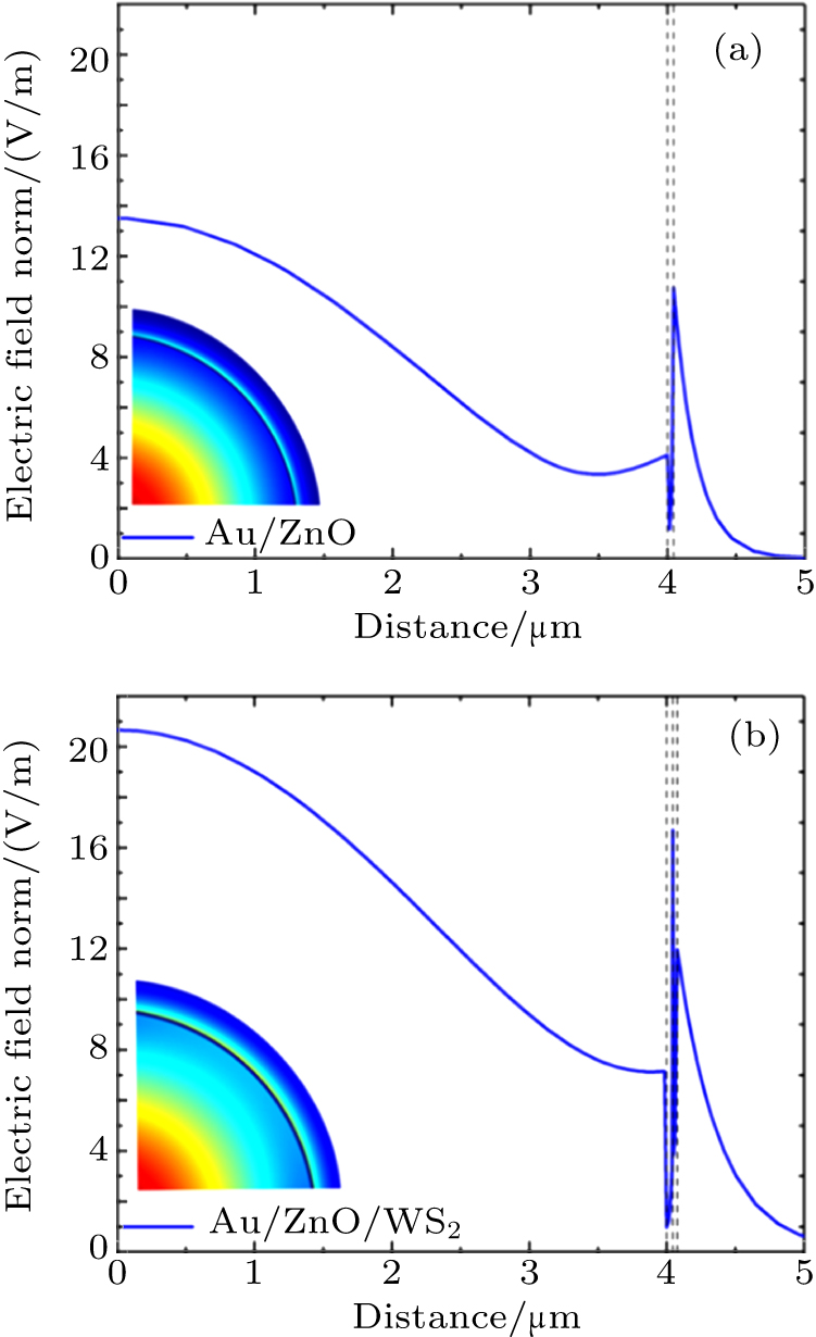
Abstract  

In this paper, we propose a theoretical model of the surface plasmon resonance-based optical fiber biosensor for detecting glucose concentration. The Au/ZnO/WS2 multilayer film is coated around optical fiber. Compared with the conventional surface plasmon resonance sensor, WS2 material can increase the sensitivity of the biosensor. The absorption capacity of WS2 is used to load glucose oxidase by forming a sensitive area to recognize glucose. Refractive index of the solution is calculated and then the concentration of the glucose can be obtained by the correspondence between refractive index and glucose concentration. The highest sensitivity of the SPR biosensor with a structure of 40-nm Au/5-nm ZnO/14 layers of WS2 is 4310 nm/RIU. The proposed WS2-based SPR fiber biosensor has a unique effect on the detection of glucose concentration. It is expected to have potential applications in future medical blood glucose concentration detection.

Keywords:  surface plasmon resonance      optical fiber      biosensor      glucose  
Received:  17 April 2020      Revised:  08 July 2020      Accepted manuscript online:  15 July 2020
Fund: the Natural Science Foundation of Jiangsu Province, China (Grant No. BK20171442), the China Postdoctoral Science Foundation (Grant No. 2018T110480), the Open Foundation of State Key Laboratory of Millimeter Waves, China (Grant No. K202003), the Open Foundation of State Key Laboratory of Luminescent Materials and Devices, China (Grant No. 2020-skllmd-03), and the Fund from the Research Center of Optical Communications Engineering & Technology, Jiangsu Province, China (Grant No. ZXF201904).
Corresponding Authors:  Corresponding author. E-mail: Liw@njupt.edu.cn   

Cite this article: 

Yun Cai(蔡云), Wei Li(李卫), Ye Feng(冯烨), Jian-Sheng Zhao(赵建胜), Gang Bai(白刚), Jie Xu(许杰), and Jin-Ze Li(李金泽)$ Sensitivity enhancement of WS2-coated SPR-based optical fiber biosensor for detecting glucose concentration 2020 Chin. Phys. B 29 110701

Fig. 1.  

Schematic diagram of SPR fiber biosensor.

Fig. 2.  

Transmission spectra of SPR biosensors with different coating materials: (a) Au, (b) Au/ZnO, and (c) Au/ZnO/WS2.

Fig. 3.  

Variations of tranmission with resonance wavelength for different values of Au film thickness.

Fig. 4.  

(a) SPR transmission spectra of proposed fiber-optic biosensor at different RI values of the analyte, and (b) variation of sensitivity with the number of WS2 layers, with inset showing the linear fits between the resonance wavelength and the RI of the analyte for various numbers of WS2 sensing layers.

Thickness of ZnO layer/nm Sensitivity/(nm/RIU) Number of WS2 layer
2 4 6 8 10 12 14 16 18
5 3750 3870 3980 4100 4180 4230 4310 4310 4290
10 3680 3710 3760 3760 3740 3680 3640 3600 3500
15 3560 3540 3490 3450 3340 3230 3150 3090 2970
20 3410 3340 3250 3160 3000 2860 2780 2720 2590
Table 1.  

Sensitivities of fiber-optic SPR biosensor with various thickness values of ZnO and WS2.

Fig. 5.  

(a) Mode field distribution of Au, Au/ZnO, and proposed Au/ZnO/WS2 SPR biosensor at sensing medium RI of 1.33, respectively, and (b) mode field distribution of proposed biosensor at the analyte RI from1.33 to 1.37 (in steps of 0.01), respectively.

Fig. 6.  

Comparison of normalized electric field versus distance curve between (a) SPR sensor with Au/ZnO structure and (b) that with Au/ZnO/WS2 structure.

Structure RI range Sensitivity/(nm/RIU) Reference
Ag/graphene 1.46–1.49 300 [40]
Au/ZnO 1.30–1.37 3161 [33]
Au/TiO2 1.333–1.403 1015.15 [41]
Au/WS2 1.333–1.360 2459.3 [30]
Au/ZnO/WS2 1.33–1.37 4310 this work
Table 2.  

Comparison of performance between proposed sensor and other sensors.

Glucose Refraction Resonance wavelength/nm
concentration/(g/l) index/(RIU) Au/ZnO Au/ZnO/WS2
1.33 603 1047
65 1.34 631 1080
149 1.35 664 1119
233 1.36 703 1165
317 1.37 750 1220
Table 3.  

Correspondence among glucose concentration, refractive index, and resonance wavelength.

Fig. 7.  

Comparison of measurement of glucose concentration between two structures.

[1]
Asif M H, Ali S M U, Nur O, Willander M, Brännmark C, Strålfors P, Englund U H, Elinder F, Danielsson B 2010 Biosens. Bioelectron. 25 2205 DOI: 10.1016/j.bios.2010.02.025
[2]
Iwasaki Y, Horiuchi T, Niwa O 2001 Anal. Chem. 73 1595 DOI: 10.1021/ac0012851
[3]
Singh S, Gupta B D 2013 Sens. Actuators B: Chem. 177 589 DOI: 10.1016/j.snb.2012.11.094
[4]
Sharma A K, Gupta B D 2007 J. Appl. Phys. 101 093111 DOI: 10.1063/1.2721779
[5]
Wu L, Chu H S, Koh W S, Li E P 2010 Opt. Express 18 14395 DOI: 10.1364/OE.18.014395
[6]
Homola J 1995 Sens. Actuators B: Chem. 29 401 DOI: 10.1016/0925-4005(95)01714-3
[7]
Lin W B, Jaffrezic-Renault N, Gagnaire A, Gagnaire H 2000 Sens. Actuators A: Phys. 84 198 DOI: 10.1016/S0924-4247(00)00345-9
[8]
Sharma A K, Gupta B D 2004 Sens. Actuators B: Chem. 100 423 DOI: 10.1016/j.snb.2004.02.013
[9]
Lee K L, Lee C W, Wang W S, Wei P K 2007 J. Bio. Opt. 12 044023 DOI: 10.1117/1.2772296
[10]
He L, Musick M D, Nicewarner S R, Salinas F G, Benkovic S J, Natan M J, Keating C D 2000 J. Am. Chem. Soc. 122 9071 DOI: 10.1021/ja001215b
[11]
Zhao J, Zhang X, Yonzon C R, Haes A J, Van Duyne R P 2006 Nanomedicine 1 219 DOI: 10.2217/17435889.1.2.219
[12]
Wu L, Guo J, Wang Q, Lu S, Dai X, Xiang Y, Fan D 2017 Sens. Actuators B: Chem. 249 542 DOI: 10.1016/j.snb.2017.04.110
[13]
Lahav A, Auslender M, Abdulhalim I 2008 Opt. Lett. 33 2539 DOI: 10.1364/OL.33.002539
[14]
Shalabney A, Abdulhalim I 2011 Laser Photon. Rev. 5 571 DOI: 10.1002/lpor.201000009
[15]
Homola J 2003 Anal. Bioanal. Chem. 377 528 DOI: 10.1007/s00216-003-2101-0
[16]
Meshginqalam B, Ahmadi M T, Ismail R, Sabatyan A 2017 Plasmon. 12 1991 DOI: 10.1007/s11468-016-0472-2
[17]
Zeng S, Baillargeat D, Ho H P, Yong K T 2014 Chem. Soc. Rev. 43 3426 DOI: 10.1039/c3cs60479a
[18]
Xu M, Liang T, Shi M, Chen H 2013 Chem. Rev. 113 3766 DOI: 10.1021/cr300263a
[19]
Eda G, Maier S A 2013 ACS Nano 7 5660 DOI: 10.1021/nn403159y
[20]
Bagdžiūnas G, Ramanavičius A 2019 Phys. Chem. Chem. Phys. 21 2968 DOI: 10.1039/C8CP07233G
[21]
Li W, Ding C, Cai Y, Liu J, Wang L, Ren Q, Xu J 2018 Sensors 18 660 DOI: 10.3390/s18020660
[22]
Li W, Dai E, Bai G, Xu J 2016 Sens. Actuators B: Chem. 237 526 DOI: 10.1016/j.snb.2016.06.133
[23]
Li W, Hu M, Ge P, Wang J, Guo Y 2014 Appl. Surf. Sci. 317 970 DOI: 10.1016/j.apsusc.2014.08.136
[24]
Ouyang Q, Zeng S, Jiang L, Hong L, Xu G, Dinh X Q, Qian J, He S, Qu J, Coquet P, Yong K T 2016 Sci. Rep. 6 28190 DOI: 10.1038/srep28190
[25]
Pandey P, Singh S P, Arya S K, Gupta V, Datta M, Singh S, Malhotra B D 2007 Langmuir. 23 3333 DOI: 10.1021/la062901c
[26]
Shan C, Yang H, Song J, Han D, Ivaska A, Niu L 2009 Anal. Chem. 81 2378 DOI: 10.1021/ac802193c
[27]
Zhang H, Meng Z, Wang Q, Zheng J 2011 Sens. Actuators B: Chem. 158 23 DOI: 10.1016/j.snb.2011.04.057
[28]
Usha S P, Shrivastav A M, Gupta B D 2016 Biosens. Bioelectron. 85 986 DOI: 10.1016/j.bios.2016.05.082
[29]
Luo Y, Chen C, Xia K, Peng S, Guan H, Tang J, Lu H, Yu J, Zhang J, Xiao Y, Chen Z 2016 Opt. Express 24 8956 DOI: 10.1364/OE.24.008956
[30]
Wang H, Zhang H, Dong J, Hu S, Zhu W, Qiu W, Lu H, Yu J, Guan H, Gao S, Li Z, Liu W, He M, Zhang J, Chen Z, Luo Y 2018 Photon. Res. 6 485 DOI: 10.1364/PRJ.6.000485
[31]
Zhu S, Gong L, Xie J, Gu Z, Zhao Y 2017 Small Methods 1 1700220 DOI: 10.1002/smtd.v1.12
[32]
Sharma A K, Gupta B D 2005 Opt. Commun. 245 159 DOI: 10.1016/j.optcom.2004.10.013
[33]
Shukla S, Sharma N K, Sajal V 2015 Sens. Actuators B: Chem. 206 463 DOI: 10.1016/j.snb.2014.09.083
[34]
Ordal M A, Long L L, Bell R J, Bell S E, Bell R R, Alexander R W, Ward C A 1983 Appl. Opt. 22 1099 DOI: 10.1364/AO.22.001099
[35]
Mishra A K, Mishra S K, Gupta B D 2015 Opt. Commun. 344 86 DOI: 10.1016/j.optcom.2015.01.043
[36]
Balevicius Z, Paulauskas A, Plikusiene I, Mikoliunaite L, Bechelany M, Popov A 2018 J. Mater. Chem. C 6 8778 DOI: 10.1039/C8TC03091J
[37]
Viter R, Balevicius Z, Chaaya A A, Baleviciute I, Tumenas S, Mikoliunaite L, Ramanavicius A, Gertnere Z, Zalesska A, Vataman V, Smyntyna V, Erts D, Miele P, Bechelany M 2015 J. Mater. Chem. C 3 6815 DOI: 10.1039/C5TC00964B
[38]
Lan C, Li C, Wang S, Yin Y, Guo H, Liu N, Liu Y 2016 RSC Adv. 6 67520 DOI: 10.1039/C6RA12643J
[39]
Yeh Y L 2008 Opt. Lasers Eng. 46 666 DOI: 10.1016/j.optlaseng.2008.04.008
[40]
Rifat A A, Mahdiraji G A, Chow D M, Shee Y G, Ahmed R, Adikan F R M 2015 Sensors 15 11499 DOI: 10.3390/s150511499
[41]
Liu K, Xue M, Jiang J, Wang T, Chang P, Liu T 2018 Opt. Commun. 410 552 DOI: 10.1016/j.optcom.2017.10.072
[1] Numerical simulation of a truncated cladding negative curvature fiber sensor based on the surface plasmon resonance effect
Zhichao Zhang(张志超), Jinhui Yuan(苑金辉), Shi Qiu(邱石), Guiyao Zhou(周桂耀), Xian Zhou(周娴), Binbin Yan(颜玢玢), Qiang Wu(吴强), Kuiru Wang(王葵如), and Xinzhu Sang(桑新柱). Chin. Phys. B, 2023, 32(3): 034208.
[2] Fiber cladding dual channel surface plasmon resonance sensor based on S-type fiber
Yong Wei(魏勇), Xiaoling Zhao(赵晓玲), Chunlan Liu(刘春兰), Rui Wang(王锐), Tianci Jiang(蒋天赐), Lingling Li(李玲玲), Chen Shi(石晨), Chunbiao Liu(刘纯彪), and Dong Zhu(竺栋). Chin. Phys. B, 2023, 32(3): 030702.
[3] Dual-channel fiber-optic surface plasmon resonance sensor with cascaded coaxial dual-waveguide D-type structure and microsphere structure
Ling-Ling Li(李玲玲), Yong Wei(魏勇), Chun-Lan Liu(刘春兰), Zhuo Ren(任卓), Ai Zhou(周爱), Zhi-Hai Liu(刘志海), and Yu Zhang(张羽). Chin. Phys. B, 2023, 32(2): 020702.
[4] Optoelectronic oscillator-based interrogation system for Michelson interferometric sensors
Ling Liu(刘玲), Xiaoyan Wu(吴小龑), Guodong Liu(刘国栋), Tigang Ning(宁提纲),Jian Xu(许建), and Haidong You(油海东). Chin. Phys. B, 2022, 31(9): 090702.
[5] A radiation-temperature coupling model of the optical fiber attenuation spectrum in the Ge/P co-doped fiber
Yong Li(李勇), Haoshi Zhang(张浩石), Xiaowei Wang(王晓伟), and Jing Jin(金靖). Chin. Phys. B, 2022, 31(7): 074211.
[6] Numerical study of a highly sensitive surface plasmon resonance sensor based on circular-lattice holey fiber
Jian-Fei Liao(廖健飞), Dao-Ming Lu(卢道明), Li-Jun Chen(陈丽军), and Tian-Ye Huang(黄田野). Chin. Phys. B, 2022, 31(6): 060701.
[7] Manipulating vector solitons with super-sech pulse shapes
Yan Zhou(周延), Keyun Zhang(张克赟), Chun Luo(罗纯), Xiaoyan Lin(林晓艳), Meisong Liao(廖梅松), Guoying Zhao(赵国营), and Yongzheng Fang(房永征). Chin. Phys. B, 2022, 31(5): 054203.
[8] High-sensitivity Bloch surface wave sensor with Fano resonance in grating-coupled multilayer structures
Daohan Ge(葛道晗), Yujie Zhou(周宇杰), Mengcheng Lv(吕梦成), Jiakang Shi(石家康), Abubakar A. Babangida, Liqiang Zhang(张立强), and Shining Zhu(祝世宁). Chin. Phys. B, 2022, 31(4): 044102.
[9] Multi-frequency focusing of microjets generated by polygonal prisms
Yu-Jing Yang(杨育静), De-Long Zhang(张德龙), and Ping-Rang Hua(华平壤). Chin. Phys. B, 2022, 31(3): 034201.
[10] Sensitivity improvement of aluminum-based far-ultraviolet nearly guided-wave surface plasmon resonance sensor
Tianqi Li(李天琦), Shujing Chen(陈淑静), and Chengyou Lin(林承友). Chin. Phys. B, 2022, 31(12): 124208.
[11] Photonic spin Hall effect and terahertz gas sensor via InSb-supported long-range surface plasmon resonance
Jie Cheng(程杰), Gaojun Wang(王高俊), Peng Dong(董鹏), Dapeng Liu(刘大鹏), Fengfeng Chi(迟逢逢), and Shengli Liu(刘胜利). Chin. Phys. B, 2022, 31(1): 014205.
[12] A multi-band and polarization-independent perfect absorber based on Dirac semimetals circles and semi-ellipses array
Zhiyou Li(李治友), Yingting Yi(易颖婷), Danyang Xu(徐丹阳), Hua Yang(杨华), Zao Yi(易早), Xifang Chen(陈喜芳), Yougen Yi(易有根), Jianguo Zhang(张建国), and Pinghui Wu(吴平辉). Chin. Phys. B, 2021, 30(9): 098102.
[13] Surface plasmon polaritons frequency-blue shift in low confinement factor excitation region
Ling-Xi Hu(胡灵犀), Zhi-Qiang He(何志强), Min Hu(胡旻), and Sheng-Gang Liu(刘盛纲). Chin. Phys. B, 2021, 30(8): 084102.
[14] Optical absorption tunability and local electric field distribution of gold-dielectric-silver three-layered cylindrical nanotube
Ye-Wan Ma(马业万), Zhao-Wang Wu(吴兆旺), Yan-Yan Jiang(江燕燕), Juan Li(李娟), Xun-Chang Yin(尹训昌), Li-Hua Zhang(章礼华), and Ming-Fang Yi(易明芳). Chin. Phys. B, 2021, 30(11): 114207.
[15] Controlled plasmon-enhanced fluorescence by spherical microcavity
Jingyi Zhao(赵静怡), Weidong Zhang(张威东), Te Wen(温特), Lulu Ye(叶璐璐), Hai Lin(林海), Jinglin Tang(唐靖霖), Qihuang Gong(龚旗煌), and Guowei Lyu(吕国伟). Chin. Phys. B, 2021, 30(11): 114215.
No Suggested Reading articles found!