Scanning the energy dissipation process of energetic materials based on excited state relaxation and vibration–vibration coupling
Wang Wen-Yan1, Sui Ning1, Zhang Li-Quan1, Wang Ying-Hui1, †, Wang Lin1, Wang Quan1, Wang Jiao1, Kang Zhi-Hui1, Yang Yan-Qiang3, Zhou Qiang2, Zhang Han-Zhuang1, ‡
Femtosecond Laser Laboratory, Key Laboratory of Physics and Technology for Advanced Batteries (Ministry of Education), College of Physics, Jilin University, Changchun 130012, China
State Key Laboratory of Superhard Materials, College of Physics, Jilin University, Changchun 130012, China
National Key Laboratory of Shock Wave and Detonation Physics, Institute of Fluid Physics, China Academy of Engineering Physics, Mianyang 621900, China


† Corresponding author. E-mail: yinghui_wang@jlu.edu.cn zhanghz@jlu.edu.cn

Project supported by the National Natural Science Foundation of China (Grant Nos. 21573094, 11274142, 11474131, 11574112, and 51502109), the National Found for Fostering Talents of Basic Science, China (Grant No. J1103202), the Science Challenging Program (Grant No. JCKY2016212A501), and China Scholarship Council (CSC) during a visit of Ning Sui (Grant No. 201706175038) to MPIA is also acknowledged.

Abstract

The energy dissipation mechanism of energetic materials (EMs) is very important for keeping safety. We choose nitrobenzene as a model of EM and employ transient absorption (TA) spectroscopy and time-resolved coherent anti-stokes Raman scattering (CARS) to clarify its energy dissipation mechanism. The TA data confirms that the excited nitrobenzene spends about 16 ps finishing the twist intramolecular charge transfer from benzene to nitro group, and dissipates its energy through the rapid vibration relaxation in the initial excited state. And then the dynamics of vibrational modes (VMs) in the ground state of nitrobenzene, which are located at 682 cm−1 (v1), 854 cm−1 (v2), 1006 cm−1 (v3), and 1023 cm−1 (v4), is scanned by CARS. It exhibits that the excess energy of nitrobenzene on the ground state would further dissipate through intramolecular vibrational redistribution based on the vibrational cooling of v1 and v2 modes, v1 and v4 modes, and v3 and v4 modes. Moreover, the vibration–vibration coupling depends not only on the energy levels of VMs, but also on the spatial position of chemical bonds relative to the VM.

1. Introduction

The interest in the energetic material has increased rapidly in the last three decades. It is one type of important functional materials and plays an important role in the fields of military affairs and civilian engineering.[1] Therefore, people have paid much attention to understand the reaction mechanism, so as to further employ the energetic materials. The previous report[2] shows that the macroscopic properties of energetic materials are much sensitive to the physical and chemical processes on the atomic or molecular scale and are also dependent on the excitation conditions. According to the special characteristics of energetic materials, the energy dissipation mechanism is very important to improve the safety and properties of energetic materials.[3] However, this process covers from femtosecond to nanosecond time scales and is much dependent on the relaxation of excited state and the damping oscillation of chemical bonds.[4] Besides the relaxation between the electronic states, the vibrational cooling and vibration–vibrational coupling also participate in the energy dissipation through intramolecular vibrational redistribution (IVR)[5] or multi-phonon up-pumping,[6] even though the energetic materials are located on the ground state. These complex dynamic processes would affect the excess energy redistribution in the energetic materials, which would induce the cleavage of the reaction bond and release chemical energy of energetic materials.[7] This finally affects the explosion rate and the reaction products. Therefore, it is very important to clarify the energy dissipation mechanism of energetic materials in detail. However, the most test technique for energetic materials could not offer the kinetic information, such as the relaxation of energy state or the vibrational cooling. Fortunately, transient absorption (TA) technique and the time-resolved coherent anti-Stokes Raman scattering (CARS) technique are two types of important ultrafast spectroscopy techniques. The former would offer the kinetic relaxation of materials in the excited state and the latter would clarify the vibration cooling and vibration–vibration coupling of chemical bonds. Therefore, we believe that the dissipation mechanism of energetic materials would be clarified by employing the TA and CARS. Among these energetic materials, the energetic materials with nitro unit are frequently used for a variety of applications.[810] So, we select nitrobenzene as a model object and discuss the energy dissipation mechanism of energetic materials by considering its energy dissipation between electronic states and that of vibrational modes together.

In this work, we study the energy dissipation process of nitrobenzene by employing transient absorption technique which detects the energy dissipation between electronic states, and time-resolved coherent anti-Stokes Raman scattering technique which tracks vibration relaxation dynamics among different vibrational modes in nitrobenzene. As a result, the relaxation between electronic states and the dynamics among vibrational energy levels are both disentangled in detail. Our results originated from TA and CARS would help us to understand the energy dissipation process of nitrobenzene at great length.

2. Experiment

Briefly, we employ a mode-lock Ti:sapphire femtosecond laser system (Coherent), which offers 2.6 mJ, 130-fs pulses at 800 nm with a repetition rate of 500 Hz. The output of the femtosecond laser beam is split into two parts by a beam splitter (9:1); the major one is frequency-doubled by a 1-mm thick beta barium borate (BBO) crystal to generate 400-nm pulses, which will be used as the pump pulse, while the minor one is focused into a 5-mm quartz cell filled with water to generate a white light continuum as the probe pulse. The excitation pulse (400 nm) is sent to a delay line and modulated by a synchronized optical chopper (Terahertz Technologies Inc., C-995) with a frequency of 250 Hz and used as the pump beam to excite the sample. The excitation spot is about 0.3 mm in diameter. The TA spectrum is carried out by a spectrometer (AvaSpec-2048 × 16). The time-resolved CARS system setup is shown in Fig. 1.[11,12] The laser pulse is split into three pulses by two beam splitters. Two of them with wave vectors k1 and k2 act as pump light. The third one act as Stokes light with wave vector kS. The three beams are spatially focused into the sample in the folded box geometry. Herein, the pump (k1 and k2) and Stokes (kS) are coming from the noncollinear optical parametric amplifiers NOPA1 and NOPA2, and their wavelengths are 560 nm and 600 nm, respectively. Through adjusting the energy difference (Δv) between the pump and Stokes laser, the Raman transition from 600 cm−1 to 1100 cm−1 may be excited resonantly. Actually, the vibrational modes of nitrobenzene would be easily detected owing to the broad femtosecond pulse in frequency domain, which would cover multi-vibration modes simultaneously. The CARS spectrum is carried out by a spectrometer (AvaSpec-2048 × 16). The dynamic signal is obtained by a photomultiplier tube (Zolix, PMTH-S1-CR131) connected to the lock-in amplifier (SR830, DSP). The sample of nitrobenzene is produced by Beijing Chemical Works and the purity is 99.5%. Nitrobenzene is hold in a 1-mm quartz cuvette.

Fig. 1. (color online) Schematic diagram of the CARS system. BS: beam splitter (50% R:50% T). Inset: the BOXCARS configuration of CARS.
3. Results and discussion

The transient absorption (TA) is employed to probe the photo-excitation relaxation process of nitrobenzene. The nitrobenzene does not emit photons after photo-excitation, suggesting that the excited nitrobenzene would release its energy non-radiatively. Meanwhile, the excess energy originated from photo-excitation would heat up the nitrobenzene.[13] Figure 2(a) exhibits the time-dependent TA spectra, where a broad negative band appears in the TA spectra. This negative band is mainly located in the visible region and out of the absorption spectrum of nitrobenzene, implying that this spectral feature should be assigned to the excited state absorption (ESA). There is a rising behavior in the frequency domain from 590 nm to 700 nm, and then its amplitude gradually decreases after 40 ps. This dynamic behavior indicates that the ESA signal should be originated from the intermediate state.[14,15] The previous work has pointed out that this intermediate state should be assigned to the twisted intramolecular charge transfer (TICT) formation in excited states,[16,17] where the benzene (donor) and nitro (acceptor) group orbitals of the system exist in an orthogonal conformation in the excited state.[18] The rising spectral feature in TA spectra should correspond to the TICT process from the benzene to the nitro unit. Figures 2(b) and 2(c) present the TA curves of nitrobenzene at different wavelengths. The temporal trace at 670 nm exhibits that a rising component appears in the initial dynamic process with lifetime (τG) of 16.1 ps, corresponding to the generation process of TICT after photo-excitation. And then this intensity monotonically decreases with time and its lifetime (τD) is about 762 ps, which is used to describe the relaxation process of TICT. The dynamics at 475 nm display a monotonous relaxation process. To retrieve characteristic time constants, this dynamic curve is fitted by tri-exponential decay and the results are given in Fig. 2(b). τVR is about 690 fs, whose dynamic process is so rapid that it should be attributed to the vibration relaxation. τG and τD in Fig. 2(b) are about 15.2 ps and 751 ps, which should be assigned to the generation and relaxation lifetimes of TICT, respectively, owing to the similar lifetime.

Fig. 2. (color online) (a) Time-dependent TA spectra of nitrobenzene, and the corresponding TA curves at (b) 475 nm and (c) 670 nm; (d) the corresponding energy dissipation mechanism among electronic states of nitrobenzene.

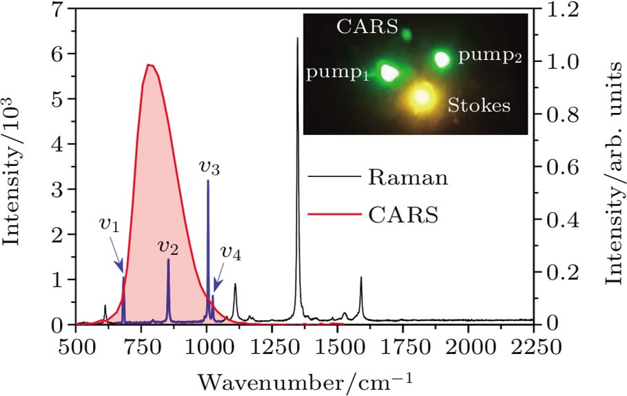
After the excited nitrobenzene relaxes on the ground state, the excess energy on the ground state would further dissipate through the vibrational dynamic behaviors involving the IVR or the vibration–vibrational coupling (VC). Therefore, the time-resolved CARS is employed to track the vibrational dynamics. Figure 3 offers the Raman spectrum and the CARS signal of nitrobenzene, where the CARS signal covers about four Raman peaks named v1, v2, v3, and v4, which correspond to 682 cm−1, 854 cm−1, 1006 cm−1, and 1023 cm−1, respectively. The corresponding motion manners are summarized in Table 1. The real picture of CARS signal and other lasers is given in the inset of Fig. 3. The direction of CARS signal is different from that of other lasers, and the detection without background would be easily obtained.

Fig. 3. (color online) Raman and CARS spectra of nitrobenzene. Note that the CARS signal (red line) overlaps with the four Raman peaks named v1 (682 cm−1), v2 (854 cm−1), v3 (1006 cm−1), and v4 (1023 cm−1). Inset: the picture of CARS signal.
Table 1.

Theoretical and experimental values of Raman shift (in unit cm−1) of nitrobenzene.[19]

.

For a better understanding of the relationship of vibration modes appearing in CARS spectrum, figure 4(a) offers the dynamic curve of total CARS signal in the direction of k1 + k2kS. Apparently, there is an obvious oscillation component signal originated from the vibration–vibration coupling, which is overlapped on the relaxation curve. Moreover, the amplitude of oscillation component decreases at the same time. In order to quantitatively analyze their components, we have to gradually extract these dynamic components from the relaxation curve of CARS signal. Note that the Gaussian curve is located near the zero time, which corresponds to the non-resonant electronic background.[20] This Gaussian component is related to the electron response to the electric filed component of light. Herein, the relaxation component follows an exponential relaxation behavior, which should correspond to the dephasing process of vibration modes. After the data fitting, it is found that the dephasing lifetime of these vibration modes is about 550 fs. The quantum beating signal appears in the relaxation curve of CARS signal as seen in Fig. 4(a), which is assigned to the coherent coupling of vibration modes originated from chemical bonds. In order to analyze this quantum beating signal, the corresponding oscillation component is extracted in Fig. 4(b). Apparently, its amplitude decreases with time, since the population in the vibrational modes would gradually decrease due to damping vibration originated from the inelastic collision with the surrounding nitrobenzene in the environment. Its lifetime is about 600 fs based on data analysis. And then, the oscillation component from 0.23 ps to 2.5 ps, which is originated from the vibration–vibration coupling, is confirmed by Fourier transform power spectra,[21] as shown in Fig. 4(c). There are three peaks at 15 cm−1, 172 cm−1, and 341 cm−1 in the frequency domain. According to the Raman spectrum of nitrobenzene in the inset of Fig. 3, it is found that these values should correspond to the energy of v4v3, v2v1, and v4v1, suggesting that the v4 mode is coupled with v3 mode, v2 mode is coupled with v1 mode, and v4 mode is coupled with v1 mode, respectively, which is summarized in Fig. 4(d). It means that the IVR would occur among these vibrational modes. According to the molecular structure of nitrobenzene, the vibration–vibration (V–V) coupling depends not only on the energy level but also on the spatial position of chemical bonds relative to the vibration mode. Moreover, the spatial position may play a distinctive role in the vibration–vibrational coupling in comparison with the energy level of the vibrational mode.

Fig. 4. (color online) (a) The dynamic curve containing the Gaussian curve (orange line); (b) the relaxation curve involving exponential decay curve (black square), oscillation signal (red circle), dephasing component (blue line), and the amplitude relaxation of oscillation signal (green line); (c) the result of the Fourier transformation of the oscillation component as shown in panels (b); (d) the coupling mechanism of v1 (682 cm−1), v2 (854 cm−1), v3 (1006 cm−1), and v4 (1023 cm−1) vibrational modes in the nitrobenzene. Inset: The total signal relaxation curve in logarithmic coordinate.
4. Conclusions

In summary, these data allow us to draw a detailed map of the relaxation pathways of excited nitrobenzene. It is found that the excited nitrobenzene has to spend about 16 ps carrying out structural twist to finish the intramolecular charge transfer from benzene to nitro group. Simultaneously, the excited nitrobenzene also dissipates its energy through the rapid vibration relaxation. After that, the nitrobenzene in the TICT state would relax to the ground state. Then, dynamics of vibrational modes in the ground state of nitrobenzene located at 682 cm−1 (v1), 854 cm−1 (v2), 1006 cm−1 (v3), and 1023 cm−1 (v4), is scanned by time-resolved CARS, which are all distributed on the benzene ring. Since the distance between the chemical bonds corresponding to these vibration modes is very close, the couplings of v4 and v3, v2, and v1, and v4 and v1 really exist, which would influence the intramolecular energy redistribution. The IVR process depends not only on the energy level but also on the spatial position of chemical bonds relative to the vibration mode.

Reference
[1] Yu Z Bernstein E R 2013 J. Phys. Chem. 117 1756
[2] Guo Y Q Greenfield M Bhattacharya A Bernstein E R 2007 J. Chem. Phys. 127 154301
[3] Zhurova E A Tsirelson V G Stash A I Yakovlev M V Pinkerton A A 2004 J. Phys. Chem. 108 20173
[4] Banerji N Cowan S Lecierc M Vauthey E Heeger A J 2010 J. Am. Chem. Soc. 132 17459
[5] Boyarkin O V Rizzo T R Perry D S 1999 J. Chem. Phys. 110 11346
[6] Dlott D D Fayer M D 1990 J. Chem. Phys. 92 3798
[7] Chen S Tolbert W A Dlott D D 1994 J. Phys. Chem. 98 7759
[8] Zander M Breymann U Dreeskamp H Koch E 1977 Z. Naturforsch. 32 1561
[9] Gerrard D L Maddams W F 1976 Appl. Spectrosc. 30 554
[10] Liu Q H Wang Y H Sui N Wang Y T Chi X C Wang Q Q Chen Y Ji W Y Zou L Zhuang H Z 2016 Sci. Rep. 6 29442
[11] Wang Y H Peng Y J He X Song Y F Yang Y Q 2009 Chin. Phys. 18 1463
[12] Namboodiri M Kazemi M M Khan T Z Materny A Kiefer J 2014 J. Am. Chem. Soc. 136 6136
[13] Takezaki M Hirota H Terazima M 1998 J. Chem. Phys. 108 4685
[14] Chi X C Ni M C Wang Y H Sui N Wang W Y Lu R Yang Y Q Ji W Y Zhang H Z 2017 J. Photoch. Photobio. 346 221
[15] Huang T H Hou J Q Kang Z H Wang Y H Lu R Zhou H P Zhao X Ma Y G Zhang H Z 2013 J. Photoch. Photobio. 261 41
[16] Sinha H K Yates K 1990 J. Chem. Phys. 93 7085
[17] Nagakura S Kojima M Maruyama Y 1964 J. Mol. Spec. 13 174
[18] Retting W 1986 Angew. Chem. Int. Ed. 25 971
[19] Zhu X M Zhang S Q Zheng X Phillips D L 2005 J. Phys. Chem. 109 3086
[20] Yu G Y Zeng Y Y Guo W C Wu H L Zhu G B Zheng Z Y Zheng X X Song Y F Yang Y Q 2017 J. Phys. Chem. 121 2565
[21] Kano H Hamaguchi H 2004 Appl. Phys. Lett. 85 4298