Measurement of electronic structure in van der Waals ferromagnet Fe5–xGeTe2
Huang Kui1, 2, 3, Li Zhenxian1, Guo Deping4, Yang Haifeng1, Li Yiwei1, 5, Liang Aiji1, 5, Wu Fan1, Xu Lixuan6, Yang Lexian6, Ji Wei4, Guo Yanfeng1, Chen Yulin1, 5, 7, †, Liu Zhongkai1, 5, ‡
ManuID:
ac5c3c
Received:2022-01-13
Copyright:
2022
More
Abstract
As a van der Waals ferromagnet with high Curie temperature, Fe5–xGeTe2 has attracted tremendous interests recently. Here, using high-resolution angle-resolved photoemission spectroscopy (ARPES), we systematically investigated the electronic structure of Fe5–xGeTe2 crystals and its temperature evolution. Our ARPES measurement reveals two types of band structures from two different terminations with slight kz evolution. Interestingly, across the ferromagnetic transition, we observed the merging of two split bands above the Curie temperature, suggesting the band splitting due to the exchange interaction within the itinerant Stoner model. Our results provide important insights into the electronic and magnetic properties of Fe5–xGeTe2 and the understanding of magnetism in a two-dimensional ferromagnetic system.
Keywords:angle-resolved photoemission spectroscopy
;
van der Waals ferromagnet
;
electronic structure
;
HuangKui, LiZhenxian, GuoDeping, YangHaifeng, LiYiwei, LiangAiji, WuFan, XuLixuan, YangLexian, JiWei, GuoYanfeng, ChenYulin, LiuZhongkai. Measurement of electronic structure in van der Waals ferromagnet Fe5–xGeTe2[J]. Chinese Physics B, 2022, 31(5): https://doi.org/10.1088/1674-1056/ac5c3c
Introduction
Recently, van der Waals (vdW) ferromagnets have attracted great research attention because they provide a versatile playground to explore the nature of the low-dimensional magnetism and a rich material basis for future applications: the vdW ferromagnets in the 2D limit exhibit unusual properties such as large magnetic anisotropy, (quantum) anomalous Hall effect, long-range magnon transport, etc.[1–6] Furthermore, their virtue of easy exfoliation into few-layer limit makes them very convenient for constructing various novel heterostructures and devices, which would find potential applications in the next-generation spintronic devices, such as spin valves, spin–orbit torque, spin field-effect transistors, spin tunneling field-effect transistors, and spin-filter magnetic tunnel junctions.
In principle, in the isotropic 2D limit, the spontaneous symmetry breaking for the formation of intrinsic long-range magnetic order is generally prohibited by strong thermal fluctuations, which is known as the Merin–Wagner theorem.[7] Nevertheless, the long-range magnetic order has been realized in a broad range of vdW ferromagnets, such as in mono- or few-layer CrI3,[1] Cr2Ge2Te6,[2] Cr2Si2Te6,[8] VSe2,[9] and MnSe2,[10] etc., owing to their intrinsic magnetocrystalline anisotropy.
Among all the vdW ferromagnets, FexGeTe2 is an intriguing material class with large intrinsic anomalous Hall effect, high Curie temperature (Tc) and highly tunable magnetic properties with different chemical composition, layer thickness, and external gating.[11–15] As for Fe3GeTe2, while the transport and optical methods have revealed 2D itinerant ferromagnetism, previous angle resolved photoemission spectroscopy (ARPES) measurement has suggested signature for non-Stoner ferromagnetism,[13] therefore the origin of the magnetic ordering as well as its interplay with the electronic structure remains mysterious and requires further investigations.
To address the issue, the investigation of new member Fe5–xGeTe2 becomes critical.[16–22] Fe5–xGeTe2 shows the highest Tc (from 270 K to 363 K depending on the Fe deficiency x) among the FexGeTe2 family, since the extra Fe layer in Fe5–xGeTe2 can considerably enhance the magnetic interaction and stabilize the long-range magnetic order. However, limited by the much less study on Fe5–xGeTe2 as compared with Fe3GeTe2, the underlying mechanisms for the room temperature Tc remain mysterious and the relation between the magnetic order and electronic structure in Fe5–xGeTe2 requires further investigations.
In this work, we systematically studied the electronic structure of Fe5–xGeTe2 using synchrotron-based ARPES and made the following discoveries. First, we have observed two kinds of band structures from two different cleaved terminations in Fe5–xGeTe2. Second, our photon energy dependence measurement shows clear periodicity of 2π/c in the electronic structure and slight variation in the band structure, which suggests the 2D nature of Fe5–xGeTe2. Finally, the temperature evolution of electronic structure of Fe5–xGeTe2 suggests the merging of two bands which are ∼ 400 meV apart across the Curie temperature. We conclude that the exchange-splitting observed in the electronic structure indicates that the itinerant Stoner model properly describes the origin of ferromagnetism in Fe5–xGeTe2. Our observation provides important insights into the nature of ferromagnetism of Fe5–xGeTe2 and other two-dimensional ferromagnetic materials.
Result and discussion
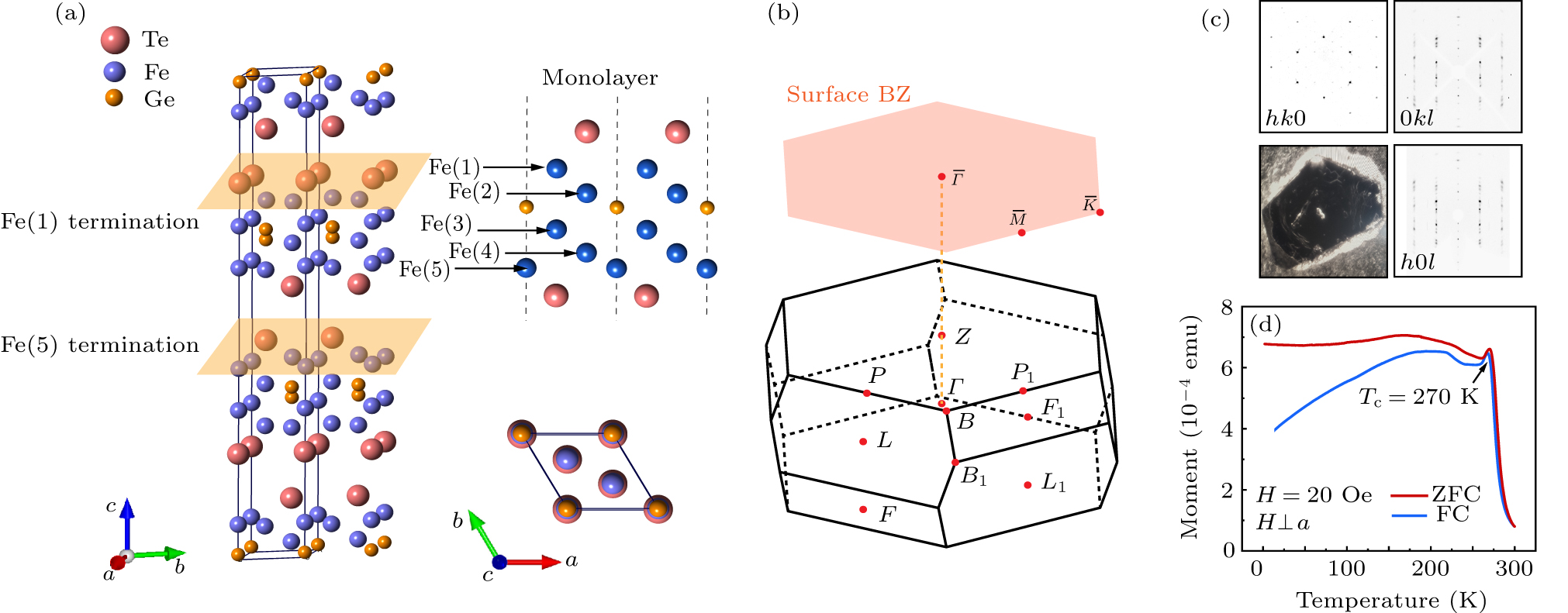
High-quality Fe5–xGeTe2 single crystals were grown from chemical vapor transport technique by using iodine as the transport agent, similar as the method described in Refs. [17,23]. The crystal structure is the rhombohedral lattice with space group (No. 166) (see Fig. 1(a) for the conventional cell). The Fe5–xGeTe2 unit cell is composed of two octuple layers bounded by the vdW force, each arranged as Te–Fe(1)–Fe(2)–Ge–Fe(3)–Fe(4)–Fe(5)–Te and Te–Fe(5)–Fe(4)–Fe(3)–Ge–Fe(2)–Fe(1)–Te, where Fe atoms occupy 5 different sublayers. Two kinds of terminations could be obtained after the cleavage along its (0001) direction with Fe(1) or Fe(5) as the top-most Fe layers (Fig. 1(a)). The corresponding Brillouin zone (BZ), its projection to the (0001) surface and the high-symmetry points are illustrated in Fig. 1(b). The samples typically appear in lamelliform as shown in Fig. 1(c), and the high quality could be verified by the x-ray diffraction (XRD) (Fig. 1(c)), from which the conventional lattice constants can be extracted as a = b = 4.05 Å, c = 29.23 Å. The ferromagnetism of the compound is characterized by the temperature-dependent magnetization measurement, which suggests a ferromagnetic phase transition occurring at Tc = 270 K, in line with the previous report.[24]
Fig. 1. Basic characterization of Fe5–xGeTe2. (a) Crystal structure of Fe5–xGeTe2. Red, blue, and orange spheres indicate Te, Fe, and Ge atoms, respectively. (b) The Brillouin zone (BZ) of Fe5–xGeTe2. High-symmetry points are labeled. (c) Image of Fe5–xGeTe2 single crystal and XRD patterns along three different crystalline directions. (d) The temperature dependent magnetization of Fe5–xGeTe2. The ferromagnetic transition temperature is marked.
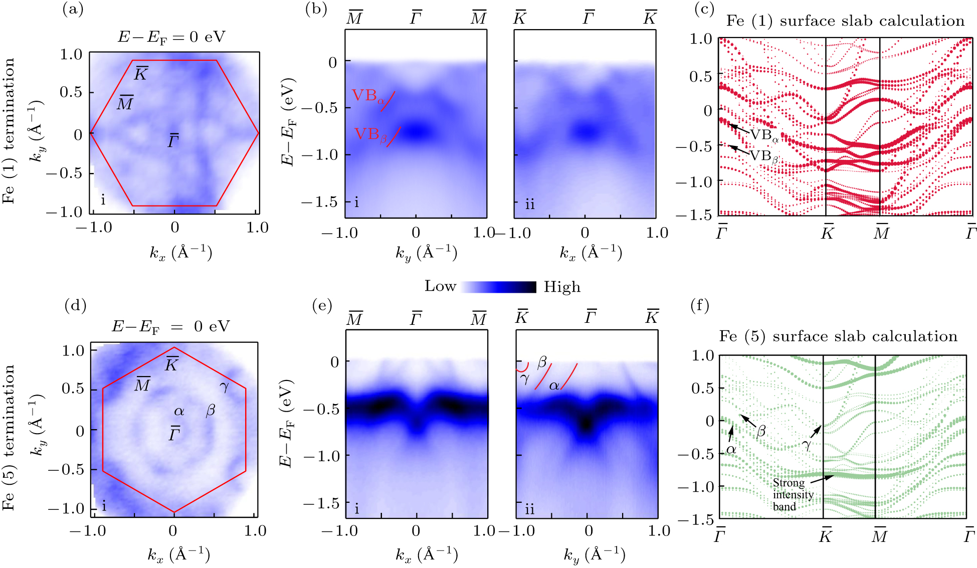
The overall electronic structure is illustrated in Fig. 2. We have observed two types of electronic structures in Fe5–xGeTe2 most likely coming from the two different terminations discussed before. Figures 2(a)–2(c)/2(d)–2(f) show the constant energy contour (CEC) in the kx–ky plane at EF, dispersion along the high symmetry –– and –– directions, and slab calculations along the high symmetry directions for Fe(1)/Fe(5) terminations, respectively. For the Fe(1) termination, we observe triangular petal-like features on the CECs at EF and characteristic features (denoted as VBα and VBβ) at higher binding energies. As for the Fe(5) termination, there are a small electron pocket near the point, a distribution of blurred spectral weight near the point and a hexagonal Fermi pocket near the point, with complex texture structure inside in Fig. 2(d), very similar to the electronic structure of Fe3GeTe2.[13] From the high-symmetry dispersions, the characteristic α and β bands correspond to the hole pockets near , while the γ band contributes to the small electron pocket near , together with one strong intensity band at the binding energy of 0.5 eV, which may come from the d orbital of excess Fe atoms. The observed characteristic bands could be captured in the slab calculations (Figs. 2(c) and 2(f)), together with other features, indicating the multiband nature of the Fe5–xGeTe2 system.
Fig. 2. Overall electronic structure of Fe5–xGeTe2 with two cleaved terminations. (a), (d) Photoemission intensity map of constant energy contours (CECs) in the kx–ky plane at EF of two cleaved terminations, respectively. The asymmetric pattern is due to the photoemission matrix element effect. (b), (e) Band dispersions along high-symmetry –– and –– of two cleaved terminations, respectively. (c), (f) Slab calculation of Fe5GeTe2 in the ferromagnetic state of two cleaved terminations, respectively. The integrated energy window of CECs is ±50 meV. The data were collected using photons with hν = 90 eV at 12 K.
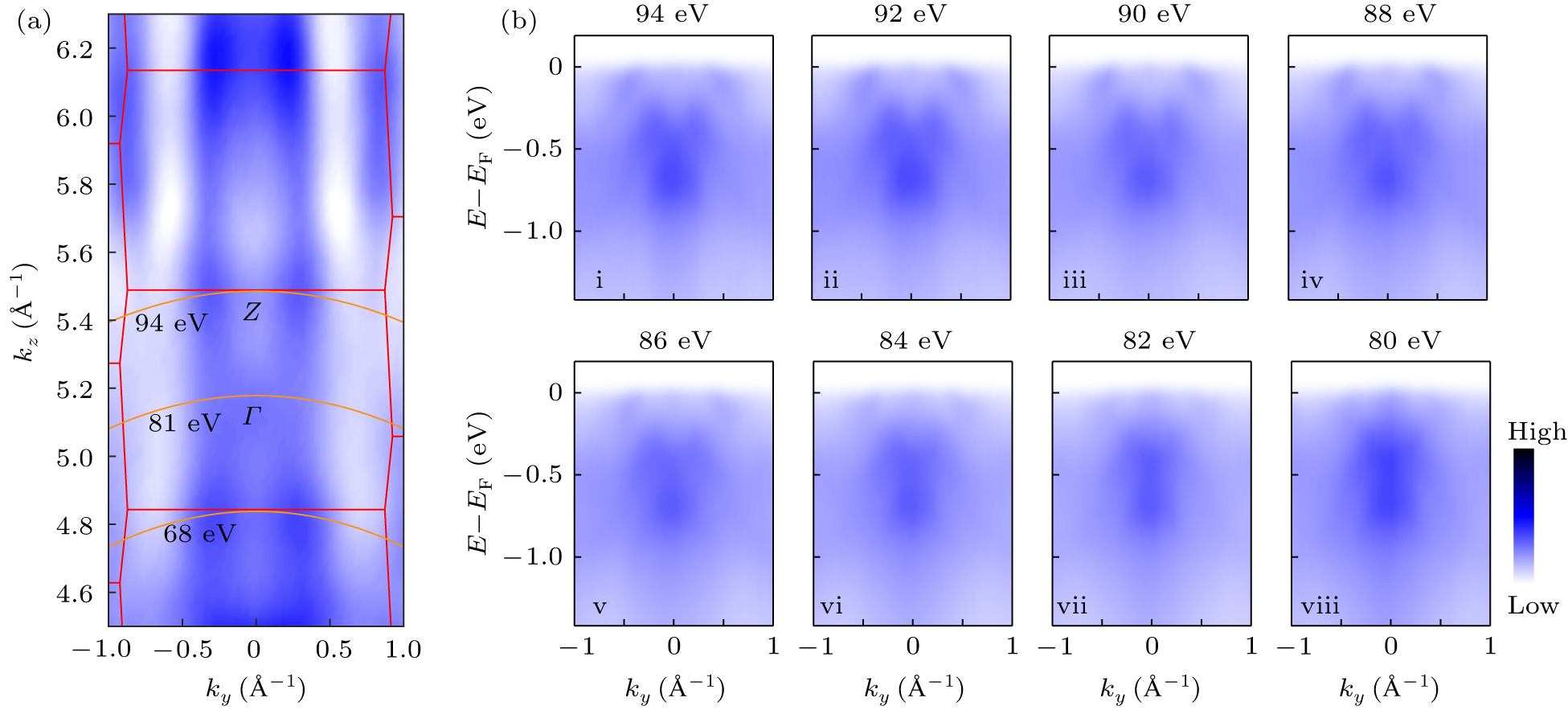
To investigate the evolution of the electronic structure along the kz direction, photon energy-dependent ARPES experiment was performed on Fe(1) termination with the result shown in Fig. 3. The CEC at 0.4 eV below EF in the ky–kz plane is plotted in Fig. 3(a), where clear periodic patterns are observed with a periodicity of 2π/c (the BZ is indicated by the red lines with high-symmetry points labeled), allowing us to determine the high symmetry points in the 3D BZ. Figure 3(b) shows the dispersion taken at photon energy from 80 eV to 94 eV along the –– direction (which spans half of a BZ). While the energy band near the Fermi surface evolves slightly with the photon energy, the bands located at higher energies show no kz evolution, which reflects the 2D nature of the Fe5–xGeTe2 compounds.
Fig. 3. The kz evolution of the electronic structure of Fe5–xGeTe2. (a) Photoemission intensity map of the CEC in the kz–ky plane at 0.4 eV below EF. Red line indicates the BZ. The orange line represents the photon energy corresponding to the high symmetry point. (b) Band dispersions along high-symmetry –– directions with different photon energy, respectively. The integrated energy window of CECs is ±50 meV. The data were collected at 12 K.
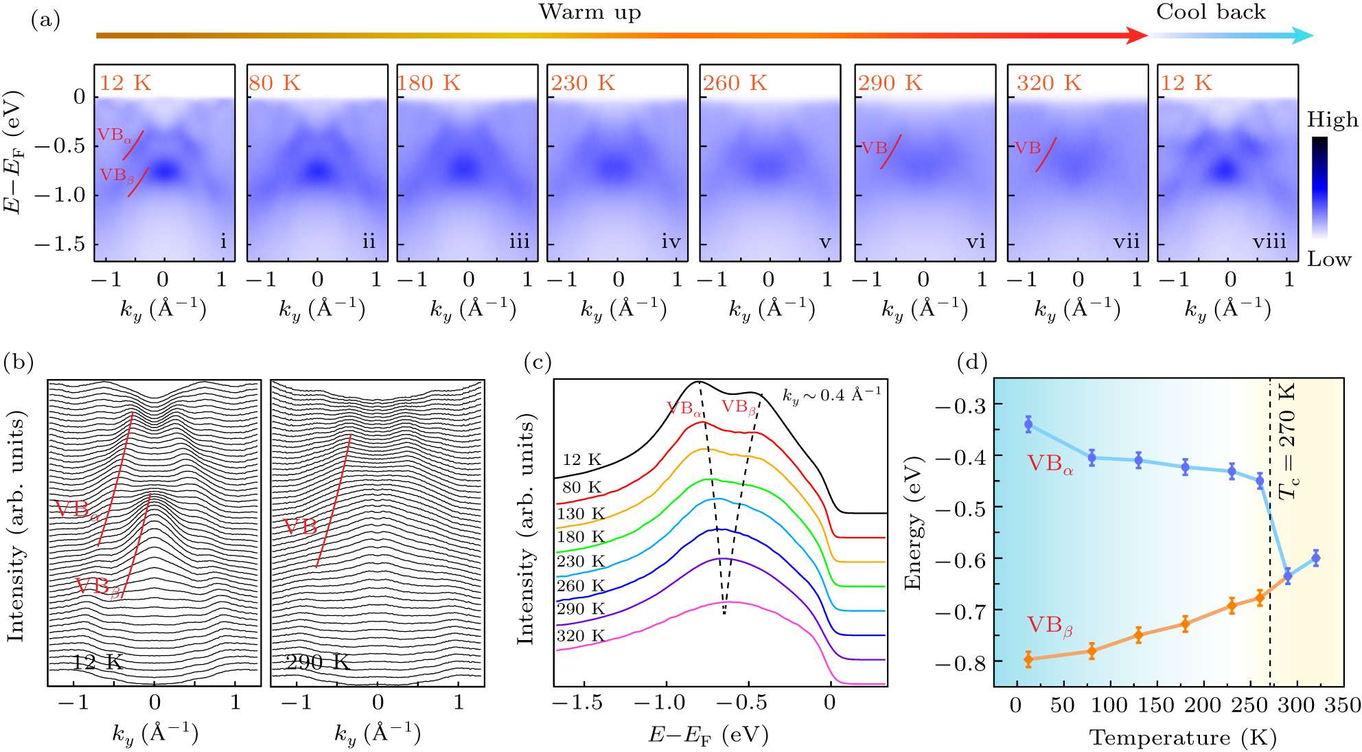
Finally, we examine the temperature evolution of the electronic bands from the ARPES data on Fe(1) termination. As shown in the plot of the original data of the ARPES intensity (Fig. 4(a)), we can see clearly that the splitting between the VBα and VBβ bands becomes smaller as the temperature increases. When the temperature is higher than the Tc (∼ 270 K), VBα and VBβ merge into one energy band (indicated by VB). When the temperature drops back below Tc, VB splits into VBα and VBβ again, ruling out the band evolution due to simple sample aging. The change of the band splitting is also clearly shown by the side-by-side comparison of stacking plot of momentum distribution curves (MDCs) between the band structures at 12 K and 290 K (Fig. 4(b)), as well as the temperature evolution of the EDCs at ky = 0.4 Å−1 (Fig. 4(c)). We extract the peak positions of the VBα and VBβ, plot their evolution at various temperatures (Fig. 4(d)) and clearly see that the band splitting closes rapidly above Tc.
Fig. 4. Temperature evolution of the electronic structure of Fe5–xGeTe2. (a) Band dispersions along high-symmetry –– directions at selected temperature. (b) The comparison of stacking plot of momentum distribution curves (MDCs) at 12 K and 290 K. The bands discussed are labeled. (c) The temperature evolution of EDCs at ky ∼ 0.4 Å−1. (d) The energy position of VBα and VBβ bands as functions of temperature. The data were collected using 90 eV photons.
In addition to the change in the splitting between the VBα and VBβ bands with increasing temperature, other complex evolutions of band structures also occur, such as the disappearing of several features near the Fermi surface (Fig. 4(a)). Although it would be quite difficult to explain the temperature evolution of the band structure of Fe5–xGeTe2 across the ferromagnetic phase transitions, especially given the reconstruction reported[25] which makes the tracing of all the bands difficult, the observed band structure evolution strongly suggests the exchange splitting of VBα and VBβ in the ferromagnetic phase. Such scenario is showing stark contrast with the band evolution of Fe3GeTe2 (and other 2D ferromagnets such as CrI3 and Cr2Ge2Te3)[13,26,27] and consistent with the itinerant Stoner model. We argue the large magnetic moment in Fe5–xGeTe2 (6.659 μB/Fe, the largest among the existing 2D ferromagnets[24]) as well as the Fe origin of the band structure observed near EF may explain the pronounced exchange splitting (∼ 400 meV) observed in Fe5–xGeTe2.
Conclusion
We have presented systematic investigation of the electronic structure of Fe5–xGeTe2. Our ARPES measurement revealed complete band structure from two different cleaved terminations. In addition, we observed a clear periodic pattern with a periodicity of 2π/c and exchange splitting of the valence band on the Fe(1) termination. Our results provide important experimental evidence of the electronic structure of Fe5–xGeTe2, and will also help to understand the magnetic origin and interaction between electrons and magnetic order of other two-dimensional ferromagnetic systems.
Method
Crystal growth
High-quality Fe5–xGeTe2 single crystals were grown by using the chemical vapor transport technique via employing iodine as the transport agent. Mixtures of high purity elements including Fe, Ge, and Te with ratio of 6 : 1 : 2 were mixed and sealed in an evacuated quartz tube and slowly heated up to 700 °C in a tubular furnace. After stayed at the temperature for 7 days, the assembly was slowly cooled down to room temperature.
Angle-resolved photoemission spectroscopy
The ARPES measurements were performed at BL03U station at Shanghai Synchrotron Radiation Facility (SSRF), China, beam line I05 of the Diamond Light Source (DLS), UK. The Fe5–xGeTe2 single crystals were first elaborately selected and well prepared in air. Then they were cleaved in situ along the (001) surface without spoiling, and measured at a 12 K sample temperature in ultrahigh vacuum with a base pressure of lower than 7 × 10⁻¹¹ Torr. Data were recorded by a VG Scienta DA30 analyzer. The angle resolution was 0.2° and the overall energy resolutions were better than 15 meV.
Density functional theory calculations
DFT calculations were performed using the generalized gradient approximation for the exchange correlation potential with a plane-wave basis and the projector augmented wave method as implemented in the Vienna ab initio simulation package (VASP).[28–30] The energy cut-off for the plane wave was set to 700 eV for structural relaxation and 500 eV for electronic structure calculations, and the k-point sampling was 15 × 15 × 1. The slab is composed of two sub-layers, and the vacuum layer is larger than 15 Å. The Fe1 atoms are above the Ge atoms in each sub-layer. The structures were fully relaxed until the residual force per atom was less than 0.001 eV/Å. In structural relaxation calculations, dispersion correction was considered in the optB86b functional for the exchange potential.[31] DFT+U method[32] was used to better describe the strong correlation system in structural relaxation and electronic structures. U = 1 eV was chosen in our calculations, and the lattice parameter a = b = 4.04 Å. Spin–orbit coupling effect and ferromagnetism were considered in electronic structure calculations.
Acknowledgments
This research used BL03U of Shanghai Synchrotron Radiation Facility and I05 of the Diamond Light Source. We also acknowledge the Analytical Instrumentation Center of ShanghaiTech University for x-ray diffraction and MPMS measurements. Z. K. Liu acknowledges the National Key R&D Program of China (Grant No. 2017YFA0305400).
... Recently, van der Waals (vdW) ferromagnets have attracted great research attention because they provide a versatile playground to explore the nature of the low-dimensional magnetism and a rich material basis for future applications: the vdW ferromagnets in the 2D limit exhibit unusual properties such as large magnetic anisotropy, (quantum) anomalous Hall effect, long-range magnon transport, etc.[1–6] Furthermore, their virtue of easy exfoliation into few-layer limit makes them very convenient for constructing various novel heterostructures and devices, which would find potential applications in the next-generation spintronic devices, such as spin valves, spin–orbit torque, spin field-effect transistors, spin tunneling field-effect transistors, and spin-filter magnetic tunnel junctions. ...
... In principle, in the isotropic 2D limit, the spontaneous symmetry breaking for the formation of intrinsic long-range magnetic order is generally prohibited by strong thermal fluctuations, which is known as the Merin–Wagner theorem.[7] Nevertheless, the long-range magnetic order has been realized in a broad range of vdW ferromagnets, such as in mono- or few-layer CrI3,[1] Cr2Ge2Te6,[2] Cr2Si2Te6,[8] VSe2,[9] and MnSe2,[10] etc., owing to their intrinsic magnetocrystalline anisotropy. ...
1
2017
... In principle, in the isotropic 2D limit, the spontaneous symmetry breaking for the formation of intrinsic long-range magnetic order is generally prohibited by strong thermal fluctuations, which is known as the Merin–Wagner theorem.[7] Nevertheless, the long-range magnetic order has been realized in a broad range of vdW ferromagnets, such as in mono- or few-layer CrI3,[1] Cr2Ge2Te6,[2] Cr2Si2Te6,[8] VSe2,[9] and MnSe2,[10] etc., owing to their intrinsic magnetocrystalline anisotropy. ...
0
2016
0
2017
0
2018
1
2018
... Recently, van der Waals (vdW) ferromagnets have attracted great research attention because they provide a versatile playground to explore the nature of the low-dimensional magnetism and a rich material basis for future applications: the vdW ferromagnets in the 2D limit exhibit unusual properties such as large magnetic anisotropy, (quantum) anomalous Hall effect, long-range magnon transport, etc.[1–6] Furthermore, their virtue of easy exfoliation into few-layer limit makes them very convenient for constructing various novel heterostructures and devices, which would find potential applications in the next-generation spintronic devices, such as spin valves, spin–orbit torque, spin field-effect transistors, spin tunneling field-effect transistors, and spin-filter magnetic tunnel junctions. ...
1
1966
... In principle, in the isotropic 2D limit, the spontaneous symmetry breaking for the formation of intrinsic long-range magnetic order is generally prohibited by strong thermal fluctuations, which is known as the Merin–Wagner theorem.[7] Nevertheless, the long-range magnetic order has been realized in a broad range of vdW ferromagnets, such as in mono- or few-layer CrI3,[1] Cr2Ge2Te6,[2] Cr2Si2Te6,[8] VSe2,[9] and MnSe2,[10] etc., owing to their intrinsic magnetocrystalline anisotropy. ...
1
2016
... In principle, in the isotropic 2D limit, the spontaneous symmetry breaking for the formation of intrinsic long-range magnetic order is generally prohibited by strong thermal fluctuations, which is known as the Merin–Wagner theorem.[7] Nevertheless, the long-range magnetic order has been realized in a broad range of vdW ferromagnets, such as in mono- or few-layer CrI3,[1] Cr2Ge2Te6,[2] Cr2Si2Te6,[8] VSe2,[9] and MnSe2,[10] etc., owing to their intrinsic magnetocrystalline anisotropy. ...
1
2018
... Among all the vdW ferromagnets, FexGeTe2 is an intriguing material class with large intrinsic anomalous Hall effect, high Curie temperature (Tc) and highly tunable magnetic properties with different chemical composition, layer thickness, and external gating.[11–15] As for Fe3GeTe2, while the transport and optical methods have revealed 2D itinerant ferromagnetism, previous angle resolved photoemission spectroscopy (ARPES) measurement has suggested signature for non-Stoner ferromagnetism,[13] therefore the origin of the magnetic ordering as well as its interplay with the electronic structure remains mysterious and requires further investigations. ...
0
2018
3
2020
... Among all the vdW ferromagnets, FexGeTe2 is an intriguing material class with large intrinsic anomalous Hall effect, high Curie temperature (Tc) and highly tunable magnetic properties with different chemical composition, layer thickness, and external gating.[11–15] As for Fe3GeTe2, while the transport and optical methods have revealed 2D itinerant ferromagnetism, previous angle resolved photoemission spectroscopy (ARPES) measurement has suggested signature for non-Stoner ferromagnetism,[13] therefore the origin of the magnetic ordering as well as its interplay with the electronic structure remains mysterious and requires further investigations. ...
... The overall electronic structure is illustrated in Fig. 2. We have observed two types of electronic structures in Fe5–xGeTe2 most likely coming from the two different terminations discussed before. Figures 2(a)–2(c)/2(d)–2(f) show the constant energy contour (CEC) in the kx–ky plane at EF, dispersion along the high symmetry –– and –– directions, and slab calculations along the high symmetry directions for Fe(1)/Fe(5) terminations, respectively. For the Fe(1) termination, we observe triangular petal-like features on the CECs at EF and characteristic features (denoted as VBα and VBβ) at higher binding energies. As for the Fe(5) termination, there are a small electron pocket near the point, a distribution of blurred spectral weight near the point and a hexagonal Fermi pocket near the point, with complex texture structure inside in Fig. 2(d), very similar to the electronic structure of Fe3GeTe2.[13] From the high-symmetry dispersions, the characteristic α and β bands correspond to the hole pockets near , while the γ band contributes to the small electron pocket near , together with one strong intensity band at the binding energy of 0.5 eV, which may come from the d orbital of excess Fe atoms. The observed characteristic bands could be captured in the slab calculations (Figs. 2(c) and 2(f)), together with other features, indicating the multiband nature of the Fe5–xGeTe2 system. ...
... In addition to the change in the splitting between the VBα and VBβ bands with increasing temperature, other complex evolutions of band structures also occur, such as the disappearing of several features near the Fermi surface (Fig. 4(a)). Although it would be quite difficult to explain the temperature evolution of the band structure of Fe5–xGeTe2 across the ferromagnetic phase transitions, especially given the reconstruction reported[25] which makes the tracing of all the bands difficult, the observed band structure evolution strongly suggests the exchange splitting of VBα and VBβ in the ferromagnetic phase. Such scenario is showing stark contrast with the band evolution of Fe3GeTe2 (and other 2D ferromagnets such as CrI3 and Cr2Ge2Te3)[13,26,27] and consistent with the itinerant Stoner model. We argue the large magnetic moment in Fe5–xGeTe2 (6.659 μB/Fe, the largest among the existing 2D ferromagnets[24]) as well as the Fe origin of the band structure observed near EF may explain the pronounced exchange splitting (∼ 400 meV) observed in Fe5–xGeTe2. ...
0
2013
1
2015
... Among all the vdW ferromagnets, FexGeTe2 is an intriguing material class with large intrinsic anomalous Hall effect, high Curie temperature (Tc) and highly tunable magnetic properties with different chemical composition, layer thickness, and external gating.[11–15] As for Fe3GeTe2, while the transport and optical methods have revealed 2D itinerant ferromagnetism, previous angle resolved photoemission spectroscopy (ARPES) measurement has suggested signature for non-Stoner ferromagnetism,[13] therefore the origin of the magnetic ordering as well as its interplay with the electronic structure remains mysterious and requires further investigations. ...
1
2018
... To address the issue, the investigation of new member Fe5–xGeTe2 becomes critical.[16–22] Fe5–xGeTe2 shows the highest Tc (from 270 K to 363 K depending on the Fe deficiency x) among the FexGeTe2 family, since the extra Fe layer in Fe5–xGeTe2 can considerably enhance the magnetic interaction and stabilize the long-range magnetic order. However, limited by the much less study on Fe5–xGeTe2 as compared with Fe3GeTe2, the underlying mechanisms for the room temperature Tc remain mysterious and the relation between the magnetic order and electronic structure in Fe5–xGeTe2 requires further investigations. ...
1
2019
... High-quality Fe5–xGeTe2 single crystals were grown from chemical vapor transport technique by using iodine as the transport agent, similar as the method described in Refs. [17,23]. The crystal structure is the rhombohedral lattice with space group (No. 166) (see Fig. 1(a) for the conventional cell). The Fe5–xGeTe2 unit cell is composed of two octuple layers bounded by the vdW force, each arranged as Te–Fe(1)–Fe(2)–Ge–Fe(3)–Fe(4)–Fe(5)–Te and Te–Fe(5)–Fe(4)–Fe(3)–Ge–Fe(2)–Fe(1)–Te, where Fe atoms occupy 5 different sublayers. Two kinds of terminations could be obtained after the cleavage along its (0001) direction with Fe(1) or Fe(5) as the top-most Fe layers (Fig. 1(a)). The corresponding Brillouin zone (BZ), its projection to the (0001) surface and the high-symmetry points are illustrated in Fig. 1(b). The samples typically appear in lamelliform as shown in Fig. 1(c), and the high quality could be verified by the x-ray diffraction (XRD) (Fig. 1(c)), from which the conventional lattice constants can be extracted as a = b = 4.05 Å, c = 29.23 Å. The ferromagnetism of the compound is characterized by the temperature-dependent magnetization measurement, which suggests a ferromagnetic phase transition occurring at Tc = 270 K, in line with the previous report.[24] ...
0
2018
0
2019
0
2018
0
2020
1
2016
... To address the issue, the investigation of new member Fe5–xGeTe2 becomes critical.[16–22] Fe5–xGeTe2 shows the highest Tc (from 270 K to 363 K depending on the Fe deficiency x) among the FexGeTe2 family, since the extra Fe layer in Fe5–xGeTe2 can considerably enhance the magnetic interaction and stabilize the long-range magnetic order. However, limited by the much less study on Fe5–xGeTe2 as compared with Fe3GeTe2, the underlying mechanisms for the room temperature Tc remain mysterious and the relation between the magnetic order and electronic structure in Fe5–xGeTe2 requires further investigations. ...
1
2019
... High-quality Fe5–xGeTe2 single crystals were grown from chemical vapor transport technique by using iodine as the transport agent, similar as the method described in Refs. [17,23]. The crystal structure is the rhombohedral lattice with space group (No. 166) (see Fig. 1(a) for the conventional cell). The Fe5–xGeTe2 unit cell is composed of two octuple layers bounded by the vdW force, each arranged as Te–Fe(1)–Fe(2)–Ge–Fe(3)–Fe(4)–Fe(5)–Te and Te–Fe(5)–Fe(4)–Fe(3)–Ge–Fe(2)–Fe(1)–Te, where Fe atoms occupy 5 different sublayers. Two kinds of terminations could be obtained after the cleavage along its (0001) direction with Fe(1) or Fe(5) as the top-most Fe layers (Fig. 1(a)). The corresponding Brillouin zone (BZ), its projection to the (0001) surface and the high-symmetry points are illustrated in Fig. 1(b). The samples typically appear in lamelliform as shown in Fig. 1(c), and the high quality could be verified by the x-ray diffraction (XRD) (Fig. 1(c)), from which the conventional lattice constants can be extracted as a = b = 4.05 Å, c = 29.23 Å. The ferromagnetism of the compound is characterized by the temperature-dependent magnetization measurement, which suggests a ferromagnetic phase transition occurring at Tc = 270 K, in line with the previous report.[24] ...
2
2020
... High-quality Fe5–xGeTe2 single crystals were grown from chemical vapor transport technique by using iodine as the transport agent, similar as the method described in Refs. [17,23]. The crystal structure is the rhombohedral lattice with space group (No. 166) (see Fig. 1(a) for the conventional cell). The Fe5–xGeTe2 unit cell is composed of two octuple layers bounded by the vdW force, each arranged as Te–Fe(1)–Fe(2)–Ge–Fe(3)–Fe(4)–Fe(5)–Te and Te–Fe(5)–Fe(4)–Fe(3)–Ge–Fe(2)–Fe(1)–Te, where Fe atoms occupy 5 different sublayers. Two kinds of terminations could be obtained after the cleavage along its (0001) direction with Fe(1) or Fe(5) as the top-most Fe layers (Fig. 1(a)). The corresponding Brillouin zone (BZ), its projection to the (0001) surface and the high-symmetry points are illustrated in Fig. 1(b). The samples typically appear in lamelliform as shown in Fig. 1(c), and the high quality could be verified by the x-ray diffraction (XRD) (Fig. 1(c)), from which the conventional lattice constants can be extracted as a = b = 4.05 Å, c = 29.23 Å. The ferromagnetism of the compound is characterized by the temperature-dependent magnetization measurement, which suggests a ferromagnetic phase transition occurring at Tc = 270 K, in line with the previous report.[24] ...
... In addition to the change in the splitting between the VBα and VBβ bands with increasing temperature, other complex evolutions of band structures also occur, such as the disappearing of several features near the Fermi surface (Fig. 4(a)). Although it would be quite difficult to explain the temperature evolution of the band structure of Fe5–xGeTe2 across the ferromagnetic phase transitions, especially given the reconstruction reported[25] which makes the tracing of all the bands difficult, the observed band structure evolution strongly suggests the exchange splitting of VBα and VBβ in the ferromagnetic phase. Such scenario is showing stark contrast with the band evolution of Fe3GeTe2 (and other 2D ferromagnets such as CrI3 and Cr2Ge2Te3)[13,26,27] and consistent with the itinerant Stoner model. We argue the large magnetic moment in Fe5–xGeTe2 (6.659 μB/Fe, the largest among the existing 2D ferromagnets[24]) as well as the Fe origin of the band structure observed near EF may explain the pronounced exchange splitting (∼ 400 meV) observed in Fe5–xGeTe2. ...
1
2021
... In addition to the change in the splitting between the VBα and VBβ bands with increasing temperature, other complex evolutions of band structures also occur, such as the disappearing of several features near the Fermi surface (Fig. 4(a)). Although it would be quite difficult to explain the temperature evolution of the band structure of Fe5–xGeTe2 across the ferromagnetic phase transitions, especially given the reconstruction reported[25] which makes the tracing of all the bands difficult, the observed band structure evolution strongly suggests the exchange splitting of VBα and VBβ in the ferromagnetic phase. Such scenario is showing stark contrast with the band evolution of Fe3GeTe2 (and other 2D ferromagnets such as CrI3 and Cr2Ge2Te3)[13,26,27] and consistent with the itinerant Stoner model. We argue the large magnetic moment in Fe5–xGeTe2 (6.659 μB/Fe, the largest among the existing 2D ferromagnets[24]) as well as the Fe origin of the band structure observed near EF may explain the pronounced exchange splitting (∼ 400 meV) observed in Fe5–xGeTe2. ...
1
2020
... In addition to the change in the splitting between the VBα and VBβ bands with increasing temperature, other complex evolutions of band structures also occur, such as the disappearing of several features near the Fermi surface (Fig. 4(a)). Although it would be quite difficult to explain the temperature evolution of the band structure of Fe5–xGeTe2 across the ferromagnetic phase transitions, especially given the reconstruction reported[25] which makes the tracing of all the bands difficult, the observed band structure evolution strongly suggests the exchange splitting of VBα and VBβ in the ferromagnetic phase. Such scenario is showing stark contrast with the band evolution of Fe3GeTe2 (and other 2D ferromagnets such as CrI3 and Cr2Ge2Te3)[13,26,27] and consistent with the itinerant Stoner model. We argue the large magnetic moment in Fe5–xGeTe2 (6.659 μB/Fe, the largest among the existing 2D ferromagnets[24]) as well as the Fe origin of the band structure observed near EF may explain the pronounced exchange splitting (∼ 400 meV) observed in Fe5–xGeTe2. ...
1
2019
... In addition to the change in the splitting between the VBα and VBβ bands with increasing temperature, other complex evolutions of band structures also occur, such as the disappearing of several features near the Fermi surface (Fig. 4(a)). Although it would be quite difficult to explain the temperature evolution of the band structure of Fe5–xGeTe2 across the ferromagnetic phase transitions, especially given the reconstruction reported[25] which makes the tracing of all the bands difficult, the observed band structure evolution strongly suggests the exchange splitting of VBα and VBβ in the ferromagnetic phase. Such scenario is showing stark contrast with the band evolution of Fe3GeTe2 (and other 2D ferromagnets such as CrI3 and Cr2Ge2Te3)[13,26,27] and consistent with the itinerant Stoner model. We argue the large magnetic moment in Fe5–xGeTe2 (6.659 μB/Fe, the largest among the existing 2D ferromagnets[24]) as well as the Fe origin of the band structure observed near EF may explain the pronounced exchange splitting (∼ 400 meV) observed in Fe5–xGeTe2. ...
1
1994
... DFT calculations were performed using the generalized gradient approximation for the exchange correlation potential with a plane-wave basis and the projector augmented wave method as implemented in the Vienna ab initio simulation package (VASP).[28–30] The energy cut-off for the plane wave was set to 700 eV for structural relaxation and 500 eV for electronic structure calculations, and the k-point sampling was 15 × 15 × 1. The slab is composed of two sub-layers, and the vacuum layer is larger than 15 Å. The Fe1 atoms are above the Ge atoms in each sub-layer. The structures were fully relaxed until the residual force per atom was less than 0.001 eV/Å. In structural relaxation calculations, dispersion correction was considered in the optB86b functional for the exchange potential.[31] DFT+U method[32] was used to better describe the strong correlation system in structural relaxation and electronic structures. U = 1 eV was chosen in our calculations, and the lattice parameter a = b = 4.04 Å. Spin–orbit coupling effect and ferromagnetism were considered in electronic structure calculations. ...
0
1999
1
1996
... DFT calculations were performed using the generalized gradient approximation for the exchange correlation potential with a plane-wave basis and the projector augmented wave method as implemented in the Vienna ab initio simulation package (VASP).[28–30] The energy cut-off for the plane wave was set to 700 eV for structural relaxation and 500 eV for electronic structure calculations, and the k-point sampling was 15 × 15 × 1. The slab is composed of two sub-layers, and the vacuum layer is larger than 15 Å. The Fe1 atoms are above the Ge atoms in each sub-layer. The structures were fully relaxed until the residual force per atom was less than 0.001 eV/Å. In structural relaxation calculations, dispersion correction was considered in the optB86b functional for the exchange potential.[31] DFT+U method[32] was used to better describe the strong correlation system in structural relaxation and electronic structures. U = 1 eV was chosen in our calculations, and the lattice parameter a = b = 4.04 Å. Spin–orbit coupling effect and ferromagnetism were considered in electronic structure calculations. ...
1
2011
... DFT calculations were performed using the generalized gradient approximation for the exchange correlation potential with a plane-wave basis and the projector augmented wave method as implemented in the Vienna ab initio simulation package (VASP).[28–30] The energy cut-off for the plane wave was set to 700 eV for structural relaxation and 500 eV for electronic structure calculations, and the k-point sampling was 15 × 15 × 1. The slab is composed of two sub-layers, and the vacuum layer is larger than 15 Å. The Fe1 atoms are above the Ge atoms in each sub-layer. The structures were fully relaxed until the residual force per atom was less than 0.001 eV/Å. In structural relaxation calculations, dispersion correction was considered in the optB86b functional for the exchange potential.[31] DFT+U method[32] was used to better describe the strong correlation system in structural relaxation and electronic structures. U = 1 eV was chosen in our calculations, and the lattice parameter a = b = 4.04 Å. Spin–orbit coupling effect and ferromagnetism were considered in electronic structure calculations. ...
1
1997
... DFT calculations were performed using the generalized gradient approximation for the exchange correlation potential with a plane-wave basis and the projector augmented wave method as implemented in the Vienna ab initio simulation package (VASP).[28–30] The energy cut-off for the plane wave was set to 700 eV for structural relaxation and 500 eV for electronic structure calculations, and the k-point sampling was 15 × 15 × 1. The slab is composed of two sub-layers, and the vacuum layer is larger than 15 Å. The Fe1 atoms are above the Ge atoms in each sub-layer. The structures were fully relaxed until the residual force per atom was less than 0.001 eV/Å. In structural relaxation calculations, dispersion correction was considered in the optB86b functional for the exchange potential.[31] DFT+U method[32] was used to better describe the strong correlation system in structural relaxation and electronic structures. U = 1 eV was chosen in our calculations, and the lattice parameter a = b = 4.04 Å. Spin–orbit coupling effect and ferromagnetism were considered in electronic structure calculations. ...