† Corresponding author. E-mail:
Project supported by the National Natural Science Foundation of China (Grant No. 61701486).
Magnetocardiography (MCG) measurement is important for investigating the cardiac biological activities. Traditionally, the extremely weak MCG signal was detected by using superconducting quantum interference devices (SQUIDs). As a room-temperature magnetic-field sensor, optically pumped magnetometer (OPM) has shown to have comparable sensitivity to that of SQUIDs, which is very suitable for biomagnetic measurements. In this paper, a synthetic gradiometer was constructed by using two OPMs under spin-exchange relaxation-free (SERF) conditions within a moderate magnetically shielded room (MSR). The magnetic noise of the OPM was measured to less than 70 fT/Hz1/2. Under a baseline of 100 mm, noise cancellation of about 30 dB was achieved. MCG was successfully measured with a signal to noise ratio (SNR) of about 37 dB. The synthetic gradiometer technique was very effective to suppress the residual environmental fields, demonstrating the OPM gradiometer technique for highly cost-effective biomagnetic measurements.
Magnetocardiography (MCG) is a contactless, non-invasive imaging technique to record the magnetic fields generated by the human heart.[1] It can visualize the myocardium activities by mapping the magnetic fields over the thorax area. The clinical trials have reported that MCG can provide a high diagnostic accuracy for many heart diseases, such as coronary artery disease (CAD), ischemic heart disease (IHD), arrhythmia, etc.[2–4] Especially, it was proved to be very valuable for the rapid screening of IHD.[5,6]
The MCG signal is extremely weak. As a highly sensitive magnetic sensor, superconducting quantum interference devices (SQUIDs) are commonly used.[7] Currently, several SQUID-based MCG systems have been commercialized and put into clinics worldwide. Despite the high sensitivity of SQUID sensors, they require liquid helium to maintain the working temperature of 4.2 K. This expensive running cost makes SQUID-based MCG systems less attractive for clinical applications. During the past few years, the optically pumped magnetometers (OPMs) have emerged to be the most attractive room-temperature magnetic sensors.[8,9] Under spin-exchange relaxation-free (SERF) conditions, OPMs can achieve a high sensitivity comparable to that of SQUIDs.[10,11]
In order to preserve a higher sensitivity, OPMs under SERF conditions must be operated under a well shielded environment. Within a magnetically shielded room (MSR) or tube, biomagnetic measurements were successfully conducted by using OPMs, such as MCG, magnetoencephyalography (MEG), etc.[12–15] However, there is still environmental disturbance although using a moderate MSR. Considering the expensive shielding costs, the popular used gradiometer techniques like SQUID systems will be a compromise method. Therefore, it will be very valuable to evaluate the gradient optically pumped measurements for the residual noise cancellation.
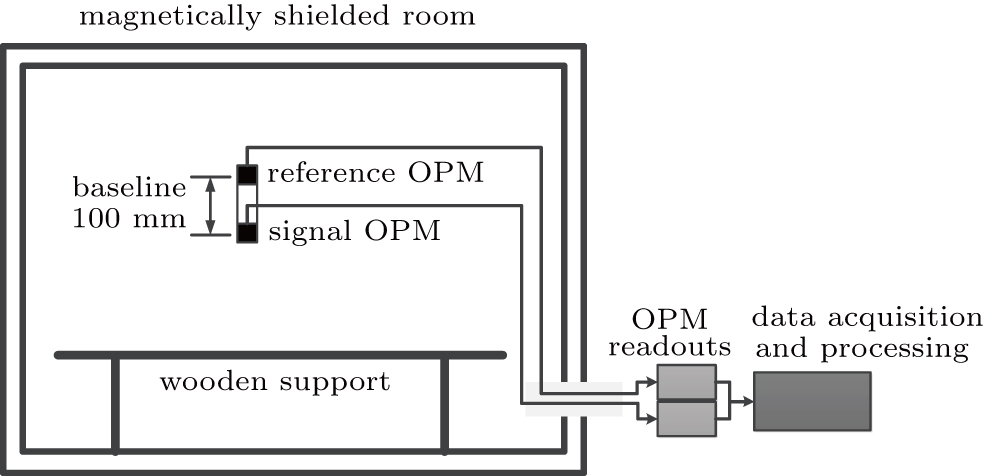
In this paper, a synthetic gradiometer method was conducted for MCG measurements. It was performed by using two individual OPMs under the SERF conditions, which were regarded as signal sensor and reference respectively. The OPM output was synthesized by using a least square algorithm with a constant subtraction coefficient. Within a moderate MSR, the gradiometer method is shown to be very effective for noise cancellation and MCG measurements, which will be applicable for developing low-cost OPM-based MCG systems.
The schematic diagram of the experimental setup is shown in Fig.
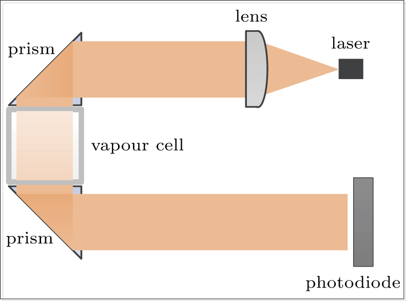
We employed commercial OPMs under SERF conditions (QuSpin Inc., Louisville, CO, USA) to make the measurements. It has a configuration of a typical noise level of 10 fT/Hz1/2, a bandwidth larger than 100 Hz, a dynamic range of about 50 nT, and a size of 12.4 mm× 16.6 mm× 24.4 mm. The basic configuration of the OPM is shown in Fig.
The analog outputs of OPMs along the vertical direction were digitized by a 24-bit 4-channel data acquisition (DAQ) system at a sample rate of 2 kHz. The raw data were pre-processed by using a 45-Hz low-pass filter to eliminate the powerline interference, which was sufficient to observe the main components of the cardiac activities. Afterwards, a synthetic gradiometer was constructed by


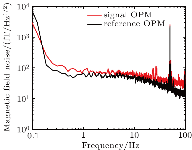
Before the MCG experiments, the power spectral density noise of the OPMs was measured firstly by using a dynamic signal analyzer, as shown in Fig.
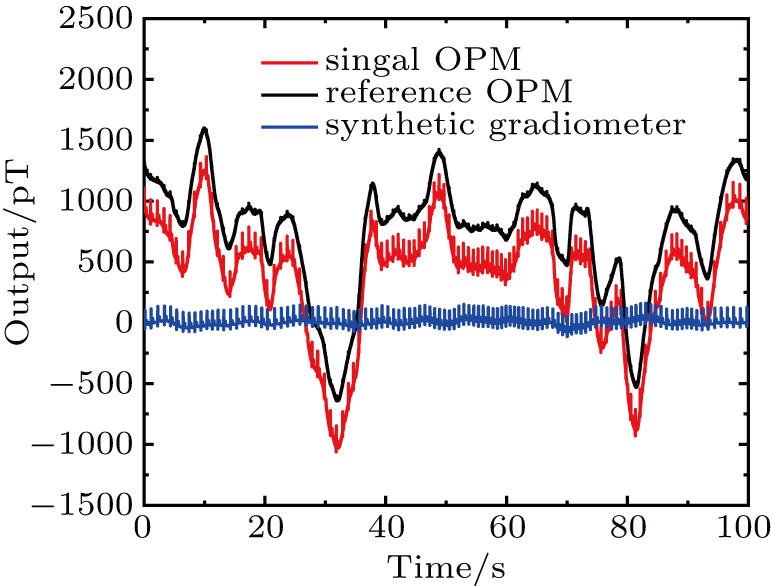
Figure
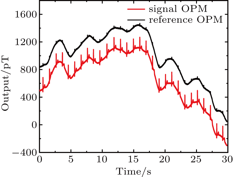 | Fig. 4. Raw output of the reference (black line) and signal (red line) OPM of an adult volunteer within a moderate MSR. |
After the low-pass filter and synthetic gradiometer process, powerline and low-frequency interferences were effectively suppressed, as shown in Fig.
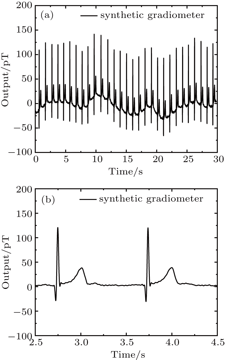 | Fig. 5. (a) Real-time output of the synthetic gradiometer; (b) two cardiac MCG cycle between 2.5 s and 4.5 s. |
In order to monitor the stability of the gradiometer output, figure
In this study, we employed two OPMs to synthesize and evaluate a gradiometer technique. Under a baseline of 100 mm, noise cancelation of about 30 dB was achieved within a moderate MSR. High quality MCG signal was measured with an SNR of about 37 dB. The synthetic gradiometer technique shows to be very effective for low-frequency noise cancellation, which will be helpful to enhance the environmental field immunity. However, low-frequency distortions still exist. The next studies will be directed to improve the system stability, such as three-axial reference compensation, detailed OPM sensor characteristics, and so on.
| [1] | |
| [2] | |
| [3] | |
| [4] | |
| [5] | |
| [6] | |
| [7] | |
| [8] | |
| [9] | |
| [10] | |
| [11] | |
| [12] | |
| [13] | |
| [14] | |
| [15] |


