Development of “Parameter space screening”-based single-wavelength anomalous diffraction phasing and structure determination pipeline
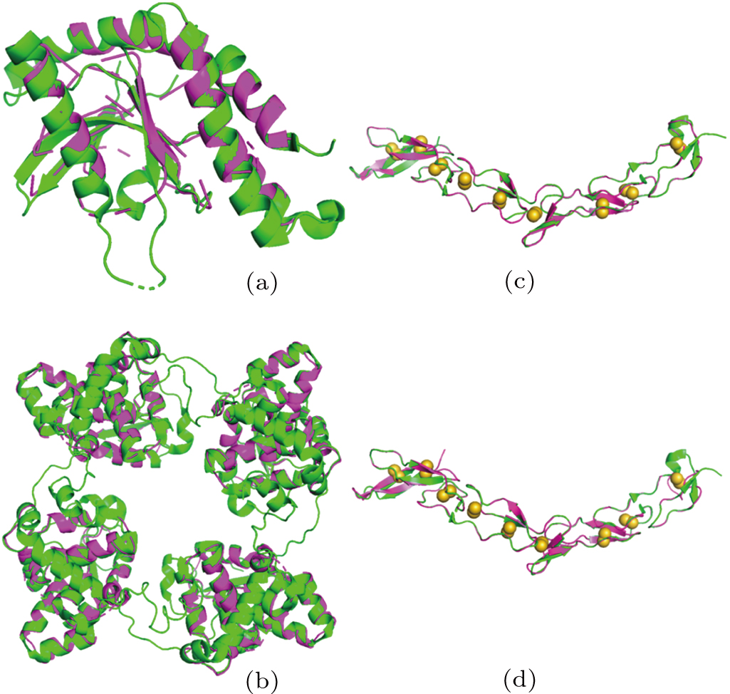
Structure comparison. Models from X2DF (in magenta) are superimposed on the structures deposited in PDB (in green), showing (a) X2DF model of T1 VS 4EF5, (b) X2DF model of T2 VS LEAV, (c) X2DF model of T3 VS 3U3S, (d) X2DF model of T4 VS 3U3P. In panels (c) and (d), disulphide bonds in deposited structures are shown as yellow spheres. There are nine disulfide bonds in 3U3S/3U3P, which belong to 18 cysteines.
