Real-space observation on standing configurations of phenylacetylene on Cu (111) by scanning probe microscopy
Qi Jing1, Gao Yi-Xuan1, Huang Li1, †, Lin Xiao1, Dong Jia-Jia2, Du Shi-Xuan1, 3, ‡, Gao Hong-Jun1, 3
       

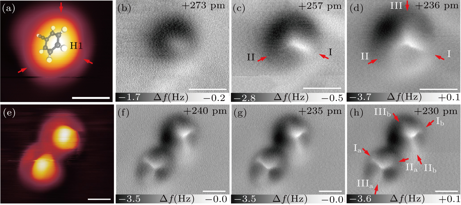
STM and constant-height nc-AFM images of PA monomer and dimer on Cu (111) substrate. (a) STM topography of PA monomer. Molecular structure of PA is superposed on top, and the topmost hydrogen atom is denoted as H1. (b)–(d) Corresponding nc-AFM images of PA monomer in Fig. 2(a) at different imaging heights. AFM imaging height refers to tunneling junction height and marked on top right of each AFM image. Red arrows indicate bright line features of PA monomer. (e) STM topography of PA dimer. (f)–(h) Corresponding nc-AFM images of PA dimer in panel (e) at different imaging heights. Red arrows indicate the bright line features of PA dimer. STM scanning parameters: VSample= −100 mV and I = 50 pA. Scale bars: 0.5 nm.