† Corresponding author. E-mail:
Project supported by the National Natural Science Foundation of China (Grant Nos. 11604155, 11604147, and 51702379), China Postdoctoral Science Foundation (Grant Nos. 2016M600428 and 2017T100386), and the Planned Projects for Postdoctoral Research Funds of China (Grant No. 1601023A).
Super-resolution optical fluctuation imaging is dependent on the blinking frequency of fluorophores. Consequently, improvement of the photoluminescence (PL) blink frequency is important. This is achieved for 3C–SiC nanocrystals (NCs) by simply increasing the excitation power. Using an excitation of 488 nm with powers of 5 μW to 50 μW, individual 3C–SiC NC always exhibits PL blinking with a short on-state sojourn time (< 0.1 s). A fast Fourier transform method is exploited to determine the PL switching frequency. It is found that the frequency of the bright state increases from 2 Hz to 20 Hz as the excitation power increases from 5 μW to 50 μW, which is explained by the Auger photonionization model.
It is well-known that many types of fluorophores, such as single molecules,[1] fluorescent proteins,[2] polymer segments,[3] semiconductor nanoparticles,[4,5] nanorods,[6] and nanowires,[7] exhibit fluorescence intermittency;[8] i.e., the photoluminescence (PL) of the fluorophores are seen to randomly flicker between emitting (“on”) and almost dark (“off”) periods under continuous excitation conditions. This phenomenon is usually called PL blinking.[9] In the case of light-emitting devices, fluorescence intermittency results in a lower quantum yield, and PL blinking is generally considered to be undesirable for conventional bioimaging or biosensing applications. This happens because fluorescence intermittency leads to a periodic loss of the of the tracking signal. Thus, much effort has been devoted to suppressing PL blinking.[10–15] However, PL blinking has revolutionized super-resolution microscopy.[16–20]
Super-resolution optical fluctuation imaging (SOFI) is a stochastic method based on PL blinking of fluorophores.[16] This method involves the higher-order statistical analysis of temporal fluorescence fluctuations recorded in a sequence of images. A fundamental assumption is that the different emitters independently switch between states in a stochastic way. The limited optical resolution of conventional fluorescent imaging techniques is attributed to the signal superposition of the fluorescence signal originating from different neighboring emitters. The n-th order cumulant (a quantity related to the n-th order correlation function) filters this signal based on its fluctuations such that only highly correlated fluctuations are retained by refining the allocation of the emitters. Specifically, the remitting signal is limited to emitters associated with a particular pixel in a detector. The fluorescence signal contribution to nearby pixels yields lower correlation values after n-th order cumulant in a nonlinear manner, which allows closely spaced emitters to be distinguished. Consequently, the width of the point spread function (PSF) is reduced by a factor of 
Silicon carbide (SiC) is an important semiconductor, which has exhibited great potential in a wide range of applications.[21–23] The PL blinking of SiC was discovered by Castelletto et al.,[24] which suggests that SiC is a potential single-photon source. This behavior was ascribed to the emission of an intrinsic defect, known as the carbon antisite–vacancy pair. Subsequently, PL blinking effects of a single 3C–SiC nanocrystal (NC) were investigated by Gan et al.,[25] and the results implied that it can be used as a fluorophore for SOFI. In this work, the excitation power density-dependent steady-state fluorescence and PL blinking of a single 3C–SiC NC are studied. It is determined that the on-state sojourn time is usually as short as several time frames under specified conditions. Moreover, as the excitation power increases, the switching frequency between emissive/dark states and on-state substantially increase.
The preparation procedure for the 3C–SiC NCs can be found elsewhere.[25] Commercial 3C–SiC powder (Alfa Aesar Co., Inc.) with a grain size of several micrometers was used as the precursor. Approximately 6.0 g of the micro-powder was etched at 100 °C for 1 h in a solution composed of 15 mL of 65-wt% nitric acid (HNO3) and 45 mL of 40-wt% hydrofluoric acid (HF). After the resulting solution was cooled, centrifugation was performed at 8000 rpm for 5 min to remove any excess acid. Subsequently, the powder obtained was washed with deionized water and dried at 70 °C in an oven. The powder was then re-dispersed using 30 mL of deionized water, followed by ultrasonic treatment for approximately 60 min. The supernatant containing 3C–SiC NCs was finally obtained by centrifugation at 8000 rpm for 10 min. Further structural characterization can also be found in a previous report.[25]
The optical absorption spectrum was obtained using a Shimadzu UV-2600 spectrometer. Ensemble PL measurements were performed on an FS5 PL spectrometer (Edinburgh Instruments). For single NC fluorescence measurements, the suspension with 3C–SiC NCs was diluted to approximately 1 nM and ultrasonically treated for one hour to individually disperse the NCs. Subsequently, a 5-μL portion was spin-coated onto a glass slide. After drying, a coverslip was placed over the glass slide and the sample was transferred and mounted on a microscope. Spatial scanning PL images were acquired using a confocal microscope (LSM780, Carl Zeiss) equipped with a spectral window of 2.9 nm and a CCD camera. The microscope setup was also used to observe and record the fluorescence trajectories of the 3C–SiC NCs.
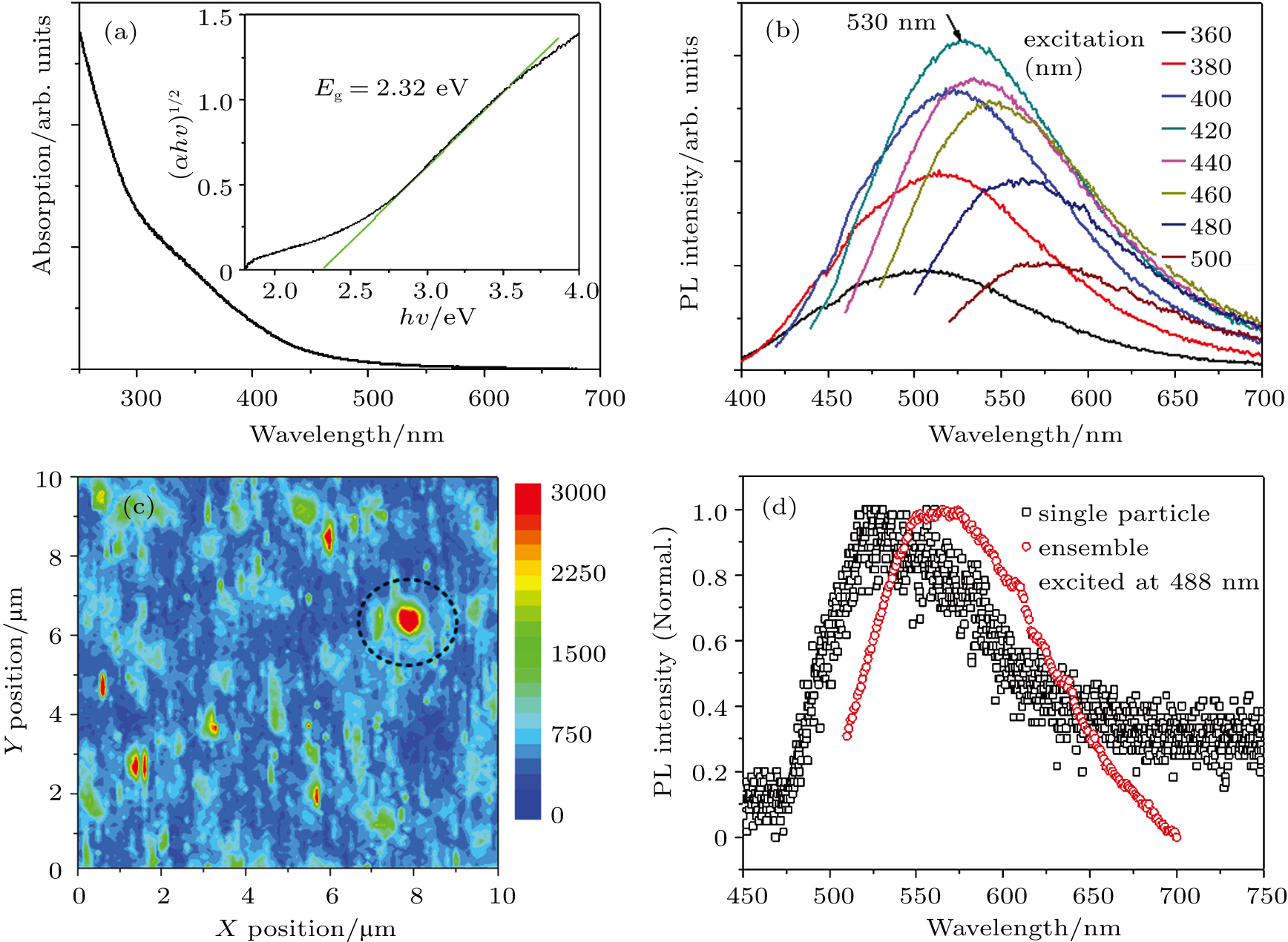
The 3C–SiC NCs with sizes ranging from 2 nm to 6 nm (most probable size of ∼3.8 nm) were synthesized by a chemical corrosion method (see previous report).[25] Most of the particles are close to or smaller than the extonic Bohr radius R (2.7 nm),[26] thus evident quantum confinement effect is expected. The bandgap of the as-prepared 3C–SiC NCs was estimated from the absorption spectrum of the corresponding aqueous solution, according to the Kubelka–Munk (KM) function. As shown in Fig.
The ensemble PL spectra of the 3C–SiC NCs dispersed in an aqueous solution is shown in Fig.
The single-particle PL spectra of the SiC NC excited at different powers were recorded. When the Ar ion laser power was increased from 10 μW to 50 μW, the PL intensity gradually increased without any apparent changes in the peak position and spectral shape (Fig.
The PL time trajectory of the single SiC NC is recorded to investigate the PL blinking. A total of 4.2 × 10⁴ frames were acquired with a time bin of 10 ms for each excitation power. All the measurements illustrated in Fig.
 | Fig. 3. (color online) PL time trajectories for the same single 3C–SiC under four different excitation powers. (a) 50, (b) 20, (c) 10, and (d) 5 μW. |
Interestingly, based on the observations from Fig.
To directly investigate the switching frequency, a fast Fourier transform (FFT) was performed on the PL time trajectories for excitation powers of 5 μW and 50 μW. The relationship between the signal magnitude and the appearance frequency is then acquired. The lowest magnitude at the highest frequency is regarded as the background (dark state) because the intensity of the dark state is equal to that of the noise and occurs most frequently. The background is marked by the shadow rectangle. Thus, the signal that appears above the shadow rectangle can be regarded as a bright state. As shown in Fig.
The photo-physics associated with the experimental observation are tentatively explained by attributing the PL blinking to Auger photon-ionization.[25,28,30] The PL is turned off by ejecting an electron to the surface trap via a double excitation. Apparently, as the excitation intensity increases, double excitation of a single NC becomes easier, which promotes Auger photon-ionization. Meanwhile, PL is switched on after the return of the ejected electron, which also requires energy transfer from the excited states. At a higher excitation intensity, the sojourn of an ejected electron at the surface, the dark state dwell time also becomes shorter. Therefore, a high excitation power facilitates the on-and-off switching process, causing the PL of 3C–SiC NCs to blink more frequently.
In summary, PL blinking at a high frequency is very important for super-resolution optical fluctuation imaging. In this work, 3C–SiC NCs with green emission was fabricated. Under 488-nm laser excitation with powers of 5 μW to 50 μW, the individual 3C–SiC NC exhibits PL blinking with a short on-state sojourn time (< 0.1 s). Conventional statistics on the occurrence of the on/off states suggest that the proportion of on states are 27.5% and 36.1% for excitation powers of 5 μW and 50 μW, respectively. Moreover, a straightforward fast Fourier transform method was developed to determine the PL switching frequency. The results verify that the frequency of the on-state increases from 2 Hz to 20 Hz, when the excitation power increases from 5 μW to 50 μW. Moreover, the PL can be modulated by various effects, such as photonic crystal and surface plasmon.[31,32] Additional approaches for controlling PL blinking will be developed in the future.
| [1] | |
| [2] | |
| [3] | |
| [4] | |
| [5] | |
| [6] | |
| [7] | |
| [8] | |
| [9] | |
| [10] | |
| [11] | |
| [12] | |
| [13] | |
| [14] | |
| [15] | |
| [16] | |
| [17] | |
| [18] | |
| [19] | |
| [20] | |
| [21] | |
| [22] | |
| [23] | |
| [24] | |
| [25] | |
| [26] | |
| [27] | |
| [28] | |
| [29] | |
| [30] | |
| [31] | |
| [32] |


