Probing conformational change of T7 RNA polymerase and DNA complex by solid-state nanopores
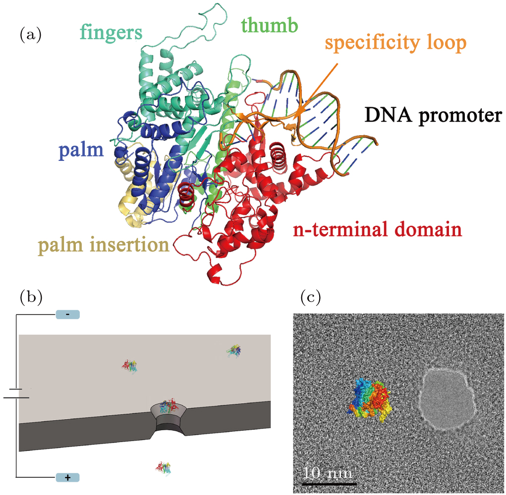
(color online) (a) PDB-based cartoon shows the structure of the T7 RNAP and complex with DNA promoter, which are colored by domain and module, with the N-terminal domain (red), the thumb (green), the palm (blue), the palm insertion module (yellow), the fingers (cyan), the specificity loop (orange). (b) Schematic of the nanopore sensor used in this work. A SiN membrane separates the electrolyte-filled space into two chambers which are connected by a nanoscale pore. An applied voltage between the electrodes generates an electrical field to drive the protein crossing the pore. (c) TEM image of a 10 nm nanopore used in this work, and a size comparison with T7 RNAP.
