Optical polarization response at gold nanosheet edges probed by scanning near-field optical microscopy
Project supported by the National Key Basic Research Program of China (Grant No. 2013CB934004) and the Fundamental Research Funds for the Central Universities, China (Grant No. YWF-13-D2-XX-14).
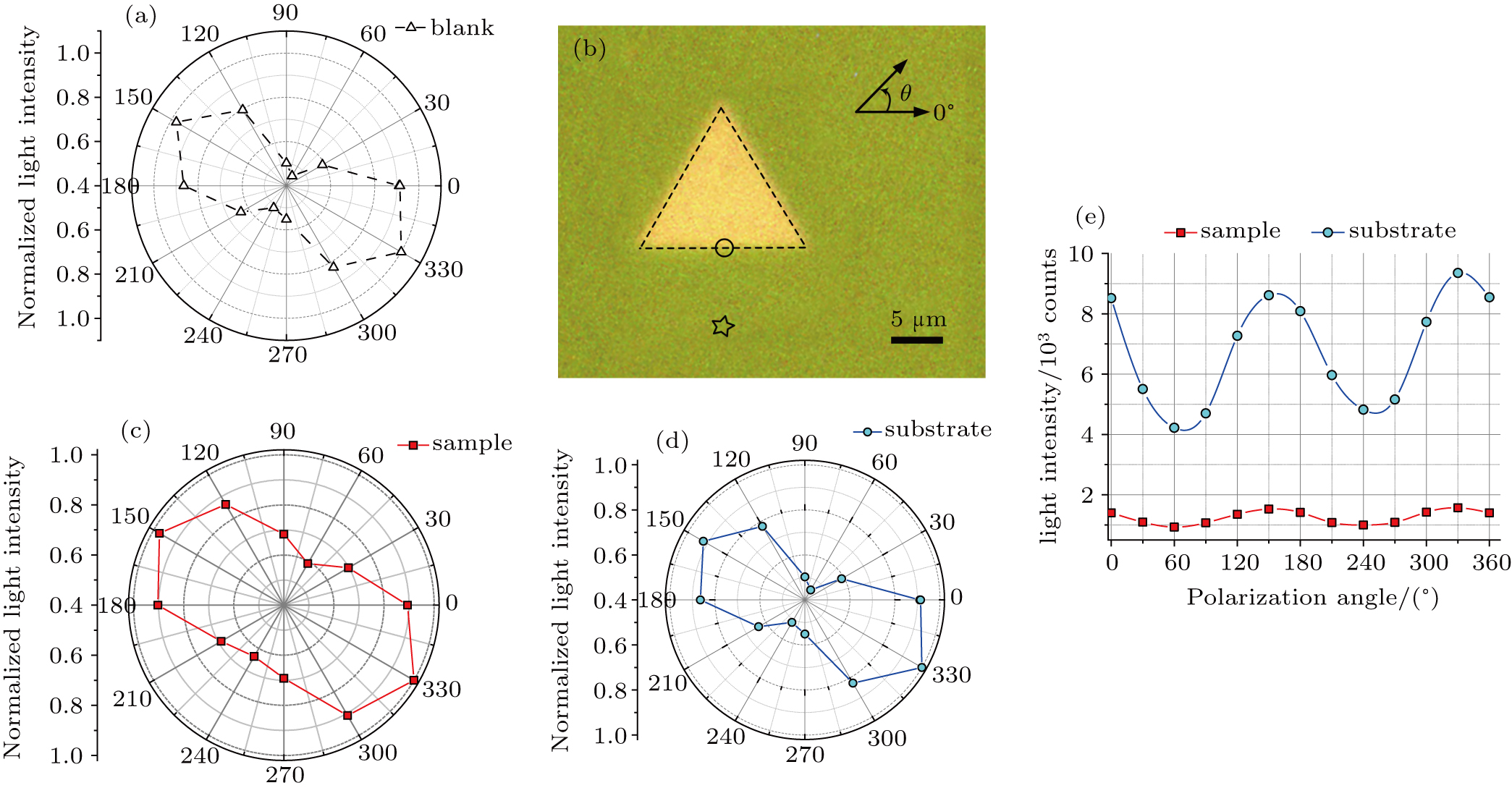
(color online) (a) Optical intensity of the tapered fiber probe varying with polarization direction of the incident light. (b) Optical image of typical gold triangle nanosheet on glass substrate. Circle and star indicate the detection positions at the edge and the glass substrate, respectively. Angle in the figure represents the polarization direction of incident light. Scale bar is 5μm long. [(c) and (d)] Normalized light intensities collected at gold edge and glass substrate, each as a function of the polarization direction in polar coordination. (e) Curves of the light intensity at the edge and the glass, each as a function of polarization angle in Cartesian coordination. The polarization angles in panels (a) and (c)–(e) are given in panel (b).
