MoS2 saturable absorber prepared by chemical vapor deposition method for nonlinear control in Q-switching fiber laser
Liu Meng-Li1, OuYang Yu-Yi1, Hou Huan-Ran1, Lei Ming1, Liu Wen-Jun1, 2, †, Wei Zhi-Yi2, ‡
State Key Laboratory of Information Photonics and Optical Communications, School of Science, Beijing University of Posts and Telecommunications, Beijing 100876, China
Beijing National Laboratory for Condensed Matter Physics, Institute of Physics, Chinese Academy of Sciences, Beijing 100190, China


† Corresponding author. E-mail: jungliu@bupt.edu.cn zywei@iphy.ac.cn

Project supported by the National Natural Science Foundation of China (Grant No. 11674036), the Beijing Youth Top-notch Talent Support Program, China (Grant No. 2017000026833ZK08), the Fund of State Key Laboratory of Information Photonics and Optical Communications, Beijing University of Posts and Telecommunications, China (Grant Nos. IPOC2016ZT04 and IPOC2017ZZ05).

Abstract

Due to the remarkable carrier mobility and nonlinear characteristic, MoS2 is considered to be a powerful competitor as an effective optical modulated material in fiber lasers. In this paper, the MoS2 films are prepared by the chemical vapor deposition method to guarantee the high quality of the crystal lattice and uniform thickness. The transfer of the films to microfiber and the operation of gold plated films ensure there is no heat-resistant damage and anti-oxidation. The modulation depth of the prepared integrated microfiber-MoS2 saturable absorber is 11.07%. When the microfiber-MoS2 saturable absorber is used as a light modulator in the Q-switching fiber laser, the stable pulse train with a pulse duration of 888 ns at 1530.9 nm is obtained. The ultimate output power and pulse energy of output pulses are 18.8 mW and 88 nJ, respectively. The signal-to-noise ratio up to 60 dB indicates the good stability of the laser. This work demonstrates that the MoS2 saturable absorber prepared by the chemical vapor deposition method can serve as an effective nonlinear control device for the Q-switching fiber laser.

1. Introduction

The saturable absorber (SA), as a natural light modulation device, is the key part of the pulsed fiber laser in Q-switching and mode-locking. Semiconductor saturable absorber mirrors (SESAMs) are the most well-researched and mature SAs, and we attribute the success in application to the controllable absorption wavelength, saturation threshold, modulation depth, and relaxation time.[1,2] However, the relatively high cost and narrow working bandwidth of SESAM have urged researchers to seek new alternatives for them. Then, the rise of graphene has meant two-dimensional (2D) materials are gradually being used more and more.[3] Some of the most representative materials that can be used as SAs have been studied in depth.[4,5] The unique zero band gap structure of graphene impels it to own the wider working wavelength than other 2D materials. The ultrafast recovery time and strong nonlinearity of graphene further prove that graphene can be a powerful candidate for SA.[69] Topological insulators (TIs) are also interesting because of their large modulation depth and nonlinearity.[1013] The superiorities of TIs in light modulation contribute to their great potential in optoelectronics applications. Black phosphorus (BPs) maintain a direct band gap whether they are a bulk or layered structure. Their band gap is adjustable from 0.3 eV to 1.5 eV, which indicates that it is easy to make a breakthrough in the application of photon and photoelectron in the near infrared band.[1418] In addition to these classic materials, some novel 2D materials have also begun to arouse interest due to their unique electronic and optical characteristics. BP quantum dots (BPQDs), which are able to combine the quantum confinement and edge effects, exhibit excellent nonlinear optical response according to previous studies.[19] Moreover, BPQDs have been successfully used in mode-locked erbium-doped fiber laser (EDFL). MXene, as another kind of new member of the 2D material family, shows good conductivity, tunable bandgap, and ability to perform ion intercalation. It has reported that MXene performs optical manipulation in photonic applications, similar to or better than graphene.[20]

In transition metal dichalcogenides (TMDs), a layer of M (Mo, W, etc.) atoms is sandwiched between two layers of X (S, Se, etc.) atoms, so the chemical formula of TMDs is generally written as MX2. The neighbor layers of TMDs are bonded with weak van der Waals force, which indicates layered nanosheets are easily separated from the sample.[2136] From previous work, the band gap of TMD varies with the number of layers, and so the consequent layer-dependent electronic and optical characteristics are attracting increasing attention.[3739] Among TMDs, MoS2 is prominent considering its performance in photoelectrons. It is reported that the nonlinear optical response of MoS2 is better than graphene.[40,41] The few-layer MoS2 unambiguously exhibits broadband absorption from previous work.[42] Furthermore, it is found that the starting Q-switching operation threshold of MoS2 is small under the same experimental conditions compared with those of the materials of the same kind.[43] The relaxation time of MoS2 is estimated at ∼30 fs as mentioned in Ref. [44], which indicates that it has potential for fabricating the ultra-fast electronic devices.

There are various methods to be chosen in the fabrication of SA. The two most representative top-down methods are mechanical exfoliation (ME)[45,46] with the aid of scotch tapes, and liquid-phase exfoliation (LPE)[47,48] getting dispersant through ultrasonication and centrifugation. These two methods are convenient and low-cost, but the layer number of prepared nanosheets is random. By comparison, the three bottom-up methods, as the common methods, have better performance in the control of the layer number and the quality of the sample, they are the pulsed laser deposition (PLD) method,[49,50] chemical vapor deposition (CVD) method,[51,52] and magnetron sputtering deposition (MSD) method,[53,54] respectively. From previous reports, the layer number of nanosheets prepared by the CVD method tends to be controlled more accurately through adjusting the reaction parameters.

Here in this paper, the MoS2 produced through the CVD method is moved to the waist of the microfiber to form MoS2 SA. Finally, the effective area of the taper fiber is fully covered with gold to prevent it from being oxidized. By means of the interaction between the evanescent field of light and the material, the SA is able to avoid some thermal damage.[55] Furthermore, the long reaction length promotes the MoS2 SA to fully exhibit the optical nonlinearity. The modulation depth (MD) and saturable intensity of MoS2 SA are measured to be 11.07% and 1.871 MW/cm2, respectively. The proposed MoS2 based Q-switching EDFL operates at 1530.9 nm. The corresponding pulse duration and maximum output power are 888 ns and 18.8 mW, respectively. The signal-to-noise ratio (SNR) (up to 60 dB) indicates the stability of the laser. The experimental results further prove the favorable optical modulation ability of MoS2 SA.

2. Preparation and characterization of MoS2 SA

The MoS2 nanosheets were grown on the ultrathin quartz plate by the CVD method. Before the synthesis, the sulfur powder of 0.5 g was placed in the reaction chamber, and the MoO3 powder of 0.5 g was placed in the heating center, which was at the downstream of sulfur powder. The ultrathin quartz plate was placed at the downstream of the heating center, which was 20 cm away from MoO3 powder. During the synthesis, the pressure of the chamber was set to be 0.1 Torr (1 Torr = 1.33322 × 10² Pa). Under the drive of Ar flow gas, MoO3 and sulfur vapors were capable of mixing and reacting fully, as the product was brought to the substrate. Because temperature is a key factor for determining the quality of the finished product, the temperature of the chamber was rising at a constant speed of 25 °C/min. The reaction chamber was heated up to 550 °C and maintained this temperature for 0.5 h, then the reaction was finished.

When the chamber cooled down to room temperature, ultrathin quartz plate attached with MoS2 film was achieved. To prevent the lattice structure of MoS2 film from destructing during transfer, the polymethyl methacrylate (PMMA) assisted method was adopted. Before the transfer, the PMMA was spin coated on a large scale over the MoS2 film. The MoS2 PMMA was able to be easily separated from ultrathin quartz plate after the open-air drying of the sample. To obtain the pure MoS2 film, the MoS2PMMA was etched in acetone. Then the pure MoS2 film was soaked in deionized water, which could not only remove the remnants, but also make the film float smoothly with the help of water tension. Finally, the MoS2 film was moved to the conical area of microfiber (SMF-28e), which was observed by using a microscope. The measured diameter of the tapered waist was 12 μm, and the effective contact length between the MoS2 film and fiber was about 2 mm. After the volatilization of superfluous deionized water, the MoS2 film closely adhered to the lateral wall of tapered fiber due to van der Waals interaction. Finally, the effective area of the microfiber was fully covered with gold to avoid rapidly oxidizing the material.

With the aid of the atomic force microscope (AFM), an important device for the nano-scale measurement, we obtained the morphology of the MoS2 surface without damaging the material. Figure 1(a) shows the particles of the material are arranged closely and orderly, which indicates the MoS2 films prepared by the CVD method are of great uniformity and crystallinity. The comparison of surface height among different regions on the MoS2 films shows the thickness of MoS2 film was 30 nm as indicated in Fig. 1(b), which corresponds to 41–42 layers in structure.

Fig. 1. (color online) (a) Surface morphology image of MoS2 film; (b) altitude difference of the surface.

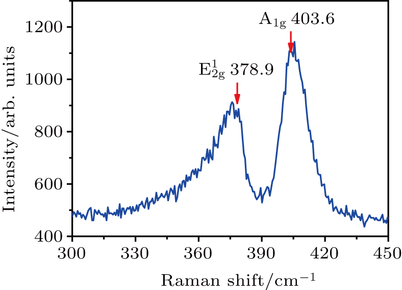
Raman spectroscopy is considered to be an effective method of determining the specific vibration modes of the material. In Fig. 2, two distinct characteristic peaks are located at 378.9 cm−1 and 403.6 cm−1. According to previous research, the mode at 378.9 cm−1 comes from the inplane vibration which belongs to Mo and S atoms, while the mode at 403.6 cm−1 comes from the out-of-plane vibration which belongs to S atoms.[5658] The frequency difference between two peaks illustrates the multilayer structure of prepared MoS2 film.

Fig. 2. (color online) Raman spectra of prepared MoS2 films.

The schematic diagram of power-dependent feature measurement is shown in Fig. 3. The light source was a mode-locked fiber laser operating at 1561 nm, of which the pulse duration and the repetition rate were 200 fs and 67 MHz. A variable fiber optic attenuator (VOA) was used to manually adjust the signal attenuation when the optical signal passes through the device. By employing OC, the optical signal could be divided into two parts: one passed through MoS2 SA to measure its saturated absorptive properties, and the other passed through SMF 28e for comparison. The optical signals at both ends were captured and recorded simultaneously by a power meter. Experimental data could be fitted by where αs, αns, and Isat are the modulation depth, nonsaturable absorption, and saturation intensity. After the fitting of experimental data in Fig. 4, the saturable intensity and MD of MoS2 SA are 1.871 MW/cm2 and 11.07%, respectively.

Fig. 3. (color online) Schematic diagram of power-dependent feature measurement.
Fig. 4. (color online) Intensity-dependent transmission of MoS2 SA, showing its nonlinear absorber characteristics.

In Table 1, the nonlinear parameters of MoS2 SAs obtained with different fabrication methods are listed to compare the advantages of CVD-MoS2. By comparison, it can be found that CVD-MoS2 generally has a large modulation depth, because the materials prepared by the CVD method have better uniformity, which is beneficial to the large modulation depth of SA. In addition, we find that the saturation intensity of CVD-MoS2 is generally small, which indicates that the laser based on CVD-MoS2 SA has a lower starting Q-switching operation threshold.

Table 1.

Nonlinear parameters of MoS2 SAs obtained with different fabrication methods.

.
3. Q-switching EDFL employed with MoS2 SA

An all-fiber ring cavity in Fig. 5 was used in the experiment. Optical devices were coupled into the laser cavity in the order corresponding to the direction of propagation of light. The pump which operated at 976 nm had the maximum allowed output power of 630 mW. The 980/1550 nm WDM successfully coupled the pump light into the ring cavity for exciting the erbium-doped fiber (EDF). The group velocity dispersion (GVD) of EDF (Liekki 110-4/125) was 12 ps2/km at 1550 nm. The intra-cavity polarization and birefringence were mainly optimized by polarization controller (PC). The primary role of an isolator (ISO) is to guarantee unidirectional optical transmission in the cavity to avoid damaging the device, caused by redundant reverse light. The MoS2 SA was placed between the ISO and WDM. The 20:80 optical coupler (OC) was adopted in the experiment, 20% of which was exported to monitor the real-time experimental data. The monitoring instruments used in the experiment mainly included the RF spectrum analyzer (Agilent E4402B), oscilloscope (Tektronix DPO3054), and optical spectrum analyzer (Yokogawa AQ6370C).

Fig. 5. (color online) Experimental illustrative diagram of Q-switching EDFL employed with MoS2 SA.

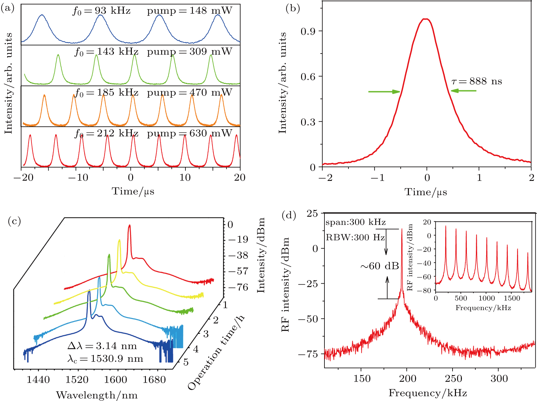
The stable Q-switching pulses occur at 147 mW with the increase of pump power. Meanwhile, the repetition rates of the output pulses with different powers are recorded and exhibited in Fig. 6(a). The tunable range of frequency is 92 kHz–212 kHz. When the pump power is increased to 630 mW, the shortest pulse duration of 888 ns is obtained as shown in Fig. 6(b). The accomplished Q-switching EDFL operates at 1530.9 nm, the corresponding spectral width is 3.14 nm. For a few-hours laser operation, the time-section data of the spectrum is shown in Fig. 6(c). The almost invariable spectral shape indicates the stability of the laser. The RF spectrum of Q-switching EDFL is provided in Fig. 6(d). The SNR of the fundamental frequency increases up to 60 dB, which indicates that the operative condition of the laser is relatively stable. Figure 6(d) shows that the downward trend of frequency multiplication is uniform, which further illustrates the stability of the laser.

Fig. 6. (color online) Experimental date of Q-switching EDFL employed with MoS2 SA, showing (a) Q-switching pulse trains at different powers, (b) single pulse waveform of Q-switching EDFL, (c) spectra at different times, and (d) RF spectrum of Q-switching EDFL.

According to the experimental monitoring data at different powers, the variation tendency of pulse duration and frequency rate with input power are shown in Fig. 7(a). In the prophase of the power growth, the pulse duration changes more sharply. However, in the later stage of power growth, the pulse duration tends to be stable. This illustrates that MoS2 SA is close to saturation at high power. With the increase of power, the repetition rate is almost increased uniformly. Similarly, we record and calculate the corresponding output power and pulse energy of the Q-switched pulse train varying with pump power in Fig. 7(b). On the whole, not only output power but also pulse energy increase evenly with input power. The ultimate output power and pulse energy are obtained to be 18.8 mW and 88 nJ, respectively. The maximum damage threshold is about 19.5 mJ/cm2.
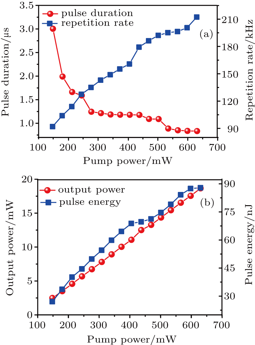
Fig. 7. (color online) (a) Variation of pulse duration and frequency rate with pump power, and (b) variation of output power and pulse energy with pump power.

Performance comparisons of Q-switched EDFL based on different SAs are shown in Table 2. By comparison, the CVD-MoS2 has certain potentials and advantages in realizing short pulse duration. Moreover, the SNR of most Q-switched EDFL is below 50 dB. The SNR of Q-switched EDFL based on CVD-MoS2 increases up to 60 dB, which indicates the stability of operation. These results show that the microfiber-MoS2 SA prepared by the CVD method is able to support stable Q-switching operation, which means it can serve as an impactful nonlinear control device for Q-switching EDFL.

Table 2.

Performance comparisons of Q-switched EDFL based on different SAs.

.
4. Conclusions

By combining the CVD method with the PMMA assisted method, the MoS2 SA based on microfiber is successfully produced. On the one hand, the films prepared by the CVD method are of good uniformity. On the other hand, the gold-plated SA based on microfiber has some advantages in preventing heat-resistant damage, and anti-oxidation. The saturate intensity and MD of MoS2 SA are 1.871 MW/cm2 and 11.07%, respectively. Moreover, the obtained MoS2 SA is successfully applied to the Q-switching EDFL. The shortest pulse duration of laser is 888 ns, and the tunable range of frequency is 92 kHz–212 kHz. The ultimate output power and pulse energy are 18.8 mW and 88 nJ, respectively. The SNR of laser increases up to 60 dB, which illustrates the stability of the operation. These results show that the microfiber-MoS2 SA prepared by the CVD method is able to support stable Q-switching pulse output, and it can serve as an impactful nonlinear control device for Q-switching EDFL.

Reference
[1] Keller U 2003 Nature 424 831
[2] Okhotnikov O Grudinin A Pessa M 2004 New J. Phys. 6 177
[3] Bao Q L Zhang H Wang Y Ni Z H Yan Y L Shen Z X Loh K P Tang D Y 2009 Adv. Funct. Mater. 19 3077
[4] Liu W J Liu M L Yin J D Chen H Lu W Fang S B Teng H Lei M Yan P G Wei Z Y 2018 Nanoscale 10 7971
[5] Liu W J Liu M L OuYang Y Y Hou H R Ma G L Lei M Wei Z Y 2018 Nanotechnology 29 174002
[6] Zhang H Bao Q L Tang D Y Zhao L M Loh K P 2009 Appl. Phys. Lett. 95 141103
[7] Sun Z P Hasan T Torrisi F Popa D Privitera G Wang F Q Bonaccorso F Basko D M Ferrari A C 2010 ACS Nano 4 803
[8] Luo A P Zhu P F Liu H Zheng X W Zhao N Liu M Cui H Luo Z C Xu W C 2014 Opt. Express 22 27019
[9] Sotor J Bogusławski J Martynkien T Mergo P Krajewska A Przewłoka A StrupiŃski W SoboŃ G 2017 Opt. Lett. 42 1592
[10] Luo Z Q Liu C Huang Y Z Wu D D Wu J Y Xu H Y Cai Z P Lin Z Q Sun L P Weng J 2014 IEEE J. Sel. Top. Quantum Electron. 20 0902708
[11] Sotor J Sobon G Grodecki K Abramski K M 2014 Appl. Phys. Lett. 104 251112
[12] Liu H Zheng X W Liu M Zhao N Luo A P Luo Z C Xu W C Zhang H Zhao C J Wen S C 2014 Opt. Express 22 6868
[13] Liu W J Pang L H Han H N Tian W L Chen H Lei M Yan P G Wei Z Y 2016 Sci. Rep. 6 19997
[14] Sotor J Sobon G Macherzynski W Paletko P Abramski K M 2015 Appl. Phys. Lett. 107 051108
[15] Hu G H Albrow-Owen T Jin X X Ali A Hu Y W Howe R C T Shehzad K Yang Z Y Zhu X K Woodward R I Wu T C Jussila H Wu J B Peng P Tan P H Sun Z P Kelleher E J R Zhang M Xu Y Hasan T 2017 Nat. Commun. 8 278
[16] Li J F Luo H Y Zhai B Lu R G Guo Z N Zhang H Liu Y 2016 Sci. Rep. 6 30361
[17] Liu S C Zhang Y N Li L Wang Y G Lv R D Wang X Chen Z D Wei L L 2018 Appl. Opt. 57 1292
[18] Yun L 2017 Opt. Express 25 32380
[19] Xu Y H Wang Z T Guo Z N Huang H Xiao Q L Zhang H 2016 Adv. Opt. Mater. 4 1223
[20] Jiang X T Liu S X Liang W Y Luo S J He Z L Ge Y Q Wang H D Cao R Zhang F Wen Q Li J Q Bao Q L Fan D Y Zhang H 2018 Laser Photon. Rev. 12 1700229
[21] Li W Y OuYang Y Y Ma G L Liu M L Liu W J 2018 Laser Phys. 28 055104
[22] Liu W J Liu M L Lei M Fang S B Wei Z Y 2018 IEEE J. Sel. Top. Quantum Electron. 24 0901005
[23] Yang C Y Li W Y Yu W T Liu M L Zhang Y J Ma G L Lei M Liu W J 2018 Nonlinear Dyn. 92 203
[24] Zhang M Howe R C T Woodward R I Kelleher E J R Torrisi F Hu G H Popov S V Taylor J R Hasan T 2015 Nano Res. 8 1522
[25] Pumera M Sofer Z Ambrosi A 2014 J. Mater. Chem. 2 8981
[26] Yan P G Liu A J Chen Y S Chen H Ruan S C Guo C Y Chen S F Li I L Yang H P Hu J G Cao G Z 2015 Opt. Mater. Express 5 479
[27] Li W Y Ma G L Yu W T Zhang Y J Liu M L Yang C Y Liu W J 2018 Chin. Phys. 27 030504
[28] Liu M L Liu W J Yan P G Fang S B Teng H Wei Z Y 2018 Chin. Opt. Lett. 16 020007
[29] Yu W T Yang C Y Liu M L Zhang Y J Liu W J 2018 Optik 159 21
[30] Huang X Zeng Z Y Zhang H 2013 Chem. Soc. Rev. 42 1934
[31] Wang J T Jiang Z K Chen H Li J R Yin J D Wang J Z He T C Yan P G Ruan S C 2017 Opt. Lett. 42 5010
[32] Liu W J Pang L H Han H N Shen Z W Lei M Teng H Wei Z Y 2016 Photon. Res. 4 111
[33] Zhang M Hu G H Hu G Q Howe R C T Chen L Zheng Z Hasan T 2015 Sci. Rep. 5 17482
[34] Liu W J Zhu Y N Liu M L Wen B Fang S B Teng H Lei M Liu L M Wei Z Y 2018 Photon. Res. 6 220
[35] Liu M L Liu W J Pang L H Teng H Fang S B Wei Z Y 2018 Opt. Commun. 406 72
[36] Liu W J Yang C Y Liu M L Yu W T Zhang Y J Lei M 2017 Phys. Rev. 96 042201
[37] Mao D She X Y Du B B Yang D X Zhang W D Song K Cui X Q Jiang B Q Peng T Zhao J L 2016 Sci. Rep. 6 23583
[38] Wang Q H Kalantar-Zadeh K Kis A Coleman J N Strano M S 2012 Nat. Nanotechnol. 7 699
[39] Britnell L Ribeiro R M Eckmann A Jalil R Belle B D Mishchenko A Kim Y J Gorbachev R V Georgiou T Morozov S V Grigorenko A N Geim A K Casiraghi C Castro Neto A H Novoselov K S 2013 Science 340 1311
[40] Du J Wang Q K Jiang G B Xu C W Zhao C J Xiang Y J Chen Y Wen S C Zhang H 2015 Sci. Rep. 4 6346
[41] Zhang H Lu S B Zheng J Du J Wen S C Tang D Y Loh K P 2014 Opt. Express 22 7249
[42] Wang S X Yu H H Zhang H J Wang A Z Zhao M W Chen Y X Mei L M Wang J Y 2014 Adv. Mater. 26 3538
[43] Chen B H Zhang X Y Wu K Wang H Wang J Chen J P 2015 Opt. Express 23 26723
[44] Wang K P Jun Wan Fan J T Lotya M O’Neill A Fox D Feng Y Y Zhang X Y Jiang B X Zhao Q Z Zhang H Z Coleman J N Zhang L Blau W J 2013 ACS Nano 7 9260
[45] Chen Y Jiang G B Chen S Q Guo Z N Yu X F Zhao C J Zhang H Bao Q L Wen S C Tang D Y Fan D Y 2015 Opt. Express 23 12823
[46] Aiub E J Steinberg D de Souza E A T Saito L A M 2017 Opt. Express 25 10546
[47] Wu K Zhang X Y Wang J Li X Chen J P 2015 Opt. Express 23 11453
[48] Koo J Park J Lee J Jhon Y M Lee J H 2016 Opt. Express 24 10575
[49] Guo B Yao Y Yan P G Xu K Liu J J Wang S G Li Yuan 2016 IEEE Photon. Technol. Lett. 28 323
[50] Liu W J Pang L H Han H N Liu M L Lei M Fang S B Teng H Wei Z Y 2017 Opt. Express 25 2950
[51] Yan P G Chen H Yin J D Xu Z H Li J R Jiang Z K Zhang W F Wang J Z Li I L Sun Z Ruan S C 2017 Nanoscale 9 1871
[52] Zhao L M Tang D Y Zhang H Wu X Bao Q L Loh K P 2010 Opt. Lett. 35 3622
[53] Chen H Chen Y S Yin J D Zhang X J Guo T Yan P G 2016 Opt. Express 24 16287
[54] Yan P G Lin R Y Ruan S C Liu A J Chen H 2015 Opt. Express 23 154
[55] Wu K Chen B H Zhang X Y Zhang S F Guo C S Li C Xiao P S Wang J Zhou L J Zou W W Chen J P 2018 Opt. Commun. 406 214
[56] Li H Zhang Q Yap C C R Tay B K Edwin T H T Olivier A Baillargeat D 2012 Adv. Funct. Mater. 22 1385
[57] Zhao Y Y Luo X Li H Zhang J Araujo P T Gan C K Wu J Zhang H Quek S Y Dresselhaus M S 2013 Nano Lett. 13 1007
[58] Lee C Yan H Brus L E Heinz T F Hone J Ryu S 2010 ACS Nano 4 2695
[59] Lou F Zhao R W He J L Jia Z T Su Xi C Wang Z W Hou J Zhang B T 2015 Photon. Res. 3 A25
[60] Luo Z C Wang F Z Liu H Liu M Tang R Luo A P Xu W C 2016 Opt. Eng. 55 081308
[61] Huang Y Z Luo Z Q Li Y Y Zhong M Xu B Che K J Xu H Y Cai Z P Peng J Weng J 2014 Opt. Express 22 25258
[62] Woodward R I Kelleher E J R Howe R C T Hu G Torrisi F Hasan T Popov S V Taylor J R 2014 Opt. Express 22 31113
[63] Luo Z Q Huang Y Z Zhong M Li Y Y Wu J Y Xu B Xu H Y Cai Z P Peng J Weng 2014 J. Lightwave Technol. 32 4077
[64] Ren J Wang S X Cheng Z C Yu H H Zhang H J Chen Y X Mei L M Wang P 2015 Opt. Express 23 5607
[65] Xia H D Li H P Lan C Y Li C Zhang X X Zhang S J Liu Y 2014 Opt. Express 22 17341
[66] Xia H D Li H P Lan C Y Li C Du J B Zhang S J Liu Y 2015 Photon. Res. 3 A92
[67] Chen H Li L Ruan S C Guo T Yan P G 2016 Opt. Eng. 55 081318
[68] Ahmad H Suthaskumar M Tiu Z C Zarei A Harun S W 2016 Opt. Laser Technol. 79 20
[69] Yu Z H Song Y R Tian J R Dou Z Y Guoyu H Y Li K X Li H W Zhang X P 2014 Opt. Express 22 11508