Phase transition and near-zero thermal expansion of Zr0.5Hf0.5VPO7*

Project supported by the National Natural Science Foundation of China (Grant Nos. 11574276, U173112, and 41401384), the Project of Shandong Provincial Higher Educational Science and Technology Program, China (Grant No. J17KB127), the Science and Technology Development Plans of Binzhou City, China (Grant Nos. 2014ZC0307 and 2015ZC0210), and Binzhou University Research Fund Project, China (Grant Nos. BZXYG1513 and BZXYG1706).

Wang Jun-Ping1, 2, Chen Qing-Dong2, Li Sai-Lei3, Ji Yan-Jun1, 2, Mu Wen-Ying2, Feng Wei-Wei2, Zeng Gao-Jie3, Liu You-Wen1, †, Liang Er-Jun3, ‡
College of Science, Nanjing University of Aeronautics and Astronautics, Nanjing 210016, China
College of Aeronautics and Engineering, Binzhou College, Binzhou 256603, China
Key Laboratory of Materials Physics of Ministry of Education, School of Physical Science and Engineering, Zhengzhou University, Zhengzhou 450052, China


† Corresponding author. E-mail: ywliu@nuaa.edu.cn ejliang@zzu.edu.cn

Project supported by the National Natural Science Foundation of China (Grant Nos. 11574276, U173112, and 41401384), the Project of Shandong Provincial Higher Educational Science and Technology Program, China (Grant No. J17KB127), the Science and Technology Development Plans of Binzhou City, China (Grant Nos. 2014ZC0307 and 2015ZC0210), and Binzhou University Research Fund Project, China (Grant Nos. BZXYG1513 and BZXYG1706).

Abstract

The Zr0.5Hf0.5VPO7 is successfully synthesized by the solid-state method with near-zero thermal expansion. Powder x-ray diffraction (XRD), Raman spectroscopy, thermal dilatometry, and scanning electron microscopy (SEM) are used to investigate the structure, the phase transition, and the coefficient of thermal expansion (CTE) of Zr0.5Hf0.5VPO7. The investigation results show that the samples are of the single cubic type with a space group of at room temperature (RT). It can be inferred that the superstructure is transformed from the 3 × 3 × 3 superstructure to the 1 × 1 × 1 ideal crystal in a temperature range between 310 K and 323 K. The CTE is measured by a dilatometer to be 0.59 × 10⁻⁶ K−1 (310 K–673 K). The values of intrinsic (XRD) and extrinsic (dilatometric) thermal expansion are both near zero. The results show that Zr0.5Hf0.5VPO7 has near-zero thermal expansion behavior over a wide temperature range.

1. Introduction

Negative thermal expansion (NTE) materials have the characteristics of heat shrinkage and cold rise, which can effectively control the expansion coefficient of material. The NTE material means it is possible to prepare composite materials each with an arbitrary expansion coefficient. The stress caused by thermal expansion and contraction is often the main cause of fatigue, performance decline, failures, and even breaking and falling off of the device. The NTE materials have extensive applications in many fields, such as optics, electronics, optical fiber communications, medicine, daily life, and many other fields. The NTE materials provide an effective way to control the thermal expansion coefficient and temperature range.[19] A series of NTE materials such as AM2O8 (A = Zr, Hf; M = W, Mo),[16] AM2O7 (A = Zr, Hf; M = V, P),[712] and A2M3O12 (A = Y, Sc, Fe; M = W, Mo) has been reported.[1319] Recently researchers investigated the materials with near-ZTE, which include Mn3Cu0.5A0.5N (A = Ni, Sn)/Cu composites,[3] Mn3Ga0.5Ge0.5N,[4] ZrW2O8/A (A = ZrO2, Al2O3, etc.) composites,[57] Zn4B6O13,[20] ZrFeMo2VO12,[21] and HfMgMo3O12.[22] The materials of MV2O7 (M = Zr, Hf) have been extensively explored to reduce the phase transition temperature.[1,7] The AM2O7 family within the cubic space group possesses the coefficient of thermal expansion (CTE) varying linearly from low positive to large negative values. The ZrV2O7 is one of the most attractive NTE materials because of its stable structure and isotropic property. However, ZrV2O7 shows a two phase transition between 350 K and 375 K. In order to further control the expansion coefficient and the phase transition temperature of ZrV2O7, researchers considered the partial substitution of V by other cations. Hisashig et al. substituted Hf4+ instead of Zr4+ solid solution of Zr1−xHfxV2O7. However, the phase transition point cannot be reduced effectively.[17] Yuan et al.[10] used Cu2+/P5+ or Fe3+/Mo5+ to replace Zr4+/V5+, but they found it was difficult to achieve a high solubility of the dopant. Many efforts have been made to design the composite materials with zero coefficient thermal expansion in a wide range of temperature. We have investigated the effects of substituting Hf4+/P5+ instead of Zr4+/V5+ and studied the effects of the phase transition and thermal expansion of ZrV2O7.

In this paper, a near-zero thermal expansion material of Zr0.5Hf0.5VPO7 is synthesized by the solid state reaction method. Thermal dilatometry, XRD, and Raman spectra are used to investigate the CTE, anisotropy, and phase transition of Zr0.5Hf0.5VPO7. It is found that the materials have the stable near-zero thermal expansion in a wide temperature range.

2. Experimental procedure

Zr0.5Hf0.5VPO7 samples were synthesized by the solid-state reaction method. The stoichiometric quantities of commercial chemicals of ZrO2 (99%), HfO2 (99%), (NH4)H2PO4 (99%), and V2O5 (99%) were ground together thoroughly with an agate mortar for 2 h. The mixed powder was heated from room temperature (RT) to 1023 K in a furnace at a rate of 5 K/min, maintained at 1023 K for 5 h, and cooled down slowly in the furnace. The sintered powder was reground, pressed into pellets with a thickness of about 10 mm and a diameter of 10 mm, and then sintered again at 1023 K for 5 h. These processes were repeated twice to implement the complete reaction. The obtained compact pellets were kept for measuring their linear CTEs.

The samples were analyzed by XRD with a PANalytical X’Pert PRO x-ray diffractometer to identify the crystalline phase. Variable-temperature x-ray powder data were collected on a Rigaku (Japan, SmartLab 3 KW) diffractometer with Cu Kα (λ = 0.15405 nm) radiation. Unit cell dimensions above the phase transition temperature were determined with software of Powder X. The relative length changes at low temperature were measured with a LINSEIS DIL L75 dilatometer at heating and cooling rates of 5 K/min. The Raman spectra of the samples were tested by a LabRAM HR Evolution Raman spectrometer. The microstructures and energy dispersive spectra of the samples were examined with a scanning electron microscope (SEM, Model Quanta 250).

3. Results
3.1. Crystal structure analysis

Figure 1 shows the Raman spectra of the solid solutions of Zr0.5Hf0.5VPO7. It is obvious that the Raman spectrum of Zr0.5Hf0.5VPO7 is very different from those of ZrV2O7 and Zr0.5Hf0.5 V2O7, which is because the radius of a P5+ ion (38 pm) is smaller than that of a V5+ ion (59 pm). Previous studies have shown that the Raman modes in the ranges of 1100 cm−1–910 cm−1, 910 cm−1–700 cm−1, 550 cm−1–450 cm−1, and 400 cm−1–300 cm−1 can be assigned to symmetric stretching (ν1), asymmetric stretching (ν3), asymmetric bending (ν4), and symmetric bending (ν2) modes of the VO4 tetrahedron, respectively. The modes below 320 cm−1 originate from lattice modes, caused by Zr atom motion, translational and vibrational modes of VO4, respectively.[1,9,23]

Fig. 1. (color online) (a) Raman spectra of (curve A) ZrV2O7, (curve B) Zr0.5Hf0.5VPO7, and (curve C) Zr0.5Hf0.5 V2O7. (b) Temperature-dependent Raman spectra of Zr0.5Hf0.5VPO7.

The most obvious change of the Raman spectrum of Zr0.5Hf0.5VPO7 is the splitting of the symmetric stretching and asymmetric stretching modes of VO4 tetrahedron around 1101 cm−1–700 cm−1 in which there are strong peaks at 1101, 1061, and 972 cm−1 belonging to the symmetric stretching vibrations (ν1 modes) of VO4. The peaks at 887, 806, and 700 cm−1 belong to asymmetric stretching (ν3 modes) of the VO4. The peaks at 524 cm−1 and 474 cm−1 are ascribed to asymmetric bending (ν4 modes). While those peaks at 403, 367, 344, 330, and 301 cm−1 originate from symmetric bending (ν2) of the VO4. The peaks at 282, 99, and 55 cm−1 originate from lattice modes, which arise from Zr atom motion, translational and librational modes of VO4.[19]

Figure 1(b) shows the temperature-dependent Raman spectra of Zr0.5Hf0.5VPO7 in a temperature range from 298 K to 673 K. The modes at about 252, 302, 402, and 700 cm−1 become weakened with temperature increasing. An obvious protrusion appears near 280 cm−1 and becomes broadened as the temperature rises. The Raman peaks do not shift and they do not change their magnitudes, either, which suggests that Zr0.5Hf0.5VPO7 keeps the cubic structure in a temperature range from 298 K to 673 K without phase transition. The results strongly suggest that Zr0.5Hf0.5VPO7 is of a cubic structure and is transformed into the 1 × 1 × 1 ideal crystal structure at RT.

Figure 2 shows the XRD patterns of the solid solutions of Zr0.5Hf0.5VPO7, Zr0.5Hf0.5V2O7, and ZrV2O7 at RT. The diffraction peaks of Zr0.5Hf0.5VO7 and Zr0.5Hf0.5V2O7 are corresponding to the primary diffraction peaks of ZrV2O7 (ICDD-JCPDS-PDFNo. 01-088-0587), which crystallizes into the cubic structure with space group . The diffraction peaks corresponding to the cubic structure recognized for ZrV2O7 were observed in Zr0.5Hf0.5V2O7.[11] However, those diffraction peaks for the superstructure almost disappear and the secondary peaks in a range of 33°–55° disappear in Zr0.5Hf0.5VPO7. All results show that the 3 × 3 × 3 superstructure of Zr0.5Hf0.5VPO7 has disappeared. However, Zr0.5Hf0.5V2O7 has such diffraction peaks for the superstructure. These results further prove that P substitution is found to be more effective to suppress the superstructure of ZrV2O7.

Fig. 2. (color online) (a) XRD patterns of (curve A) Zr0.5Hf0.5VPO7, (curve B) Zr0.5Hf0.5 V2O7, (curve C) ZrV2O7. (b) Magnified part of patterns in panel (a) (symbol ♦ indicates the diffraction peaks of superstructures).

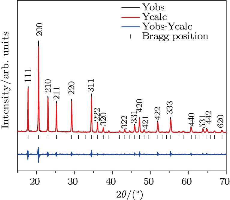
In order to confirm the crystal structure, the diffraction data are analyzed by the Rietveld and Le Bail methods in FullProf. The XRD data are refined in with the cubic structure of ZrV2O7 as a starting model. Figure 3 and Table 1 show the Rietveld analysis results of Zr0.5Hf0.5VPO7. It is shown that the Zr0.5Hf0.5VPO7 has a cubic structure with the space group (Rp = 16.7, Rwp = 16.3, Rexp = 5.09, x2 = Rwp/Rexp = 3.2). The cell parameters are calculated to be a = b = c = 8.5132 Å and α = β = γ = 90° at RT. The refined results further indicate that Zr0.5Hf0.5VPO7 adopts a cubic structure with the space group (Z = 108).

Fig. 3. (color online) Results of the Rietveld analysis of the XRD pattern for Zr0.5Hf0.5VPO7 at room temperature (Rp = 14.1, Rwp = 13.7, Rexp = 5.48, x2 = Rwp/Rexp = 2.5).
Table 1.

Detailed XRD refinement results of Zr0.5Hf0.5VPO7.

.
3.2. Thermal expansion properties

The near-ZTE property of Zr0.5Hf0.5VPO7 is investigated with a dilatometer. Figure 4 shows the variations of the relative length of Zr0.5Hf0.5VPO7 and ZrV2O7 with temperatures. The Zr0.5Hf0.5VPO7 presents an excellent stable zero thermal expansion property in the process of heating and cooling after undergoing a transition from superstructure to an ideal structure. The CET of Zr0.5Hf0.5VPO7 is obtained to be 0.59 × 10⁻⁶ K−1 in a temperature from 310 K to 673 K. This result suggests that the Zr0.5Hf0.5VPO7 has a better low thermal expansion property in a relatively low temperature range. It can also be obviously seen that there is no abrupt change below 310 K during the linear thermal expansion of the samples, indicating that the 3 × 3 × 3 superstructure has disappeared and no abrupt thermal expansion is observed, either.

Fig. 4. (color online) Relative length changes of (curve a) ZrV2O7 and (curve b) Zr0.5Hf0.5VPO7 with temperature increasing from 200 K to 673 K and temperature decreasing from 673 K to 310 K.

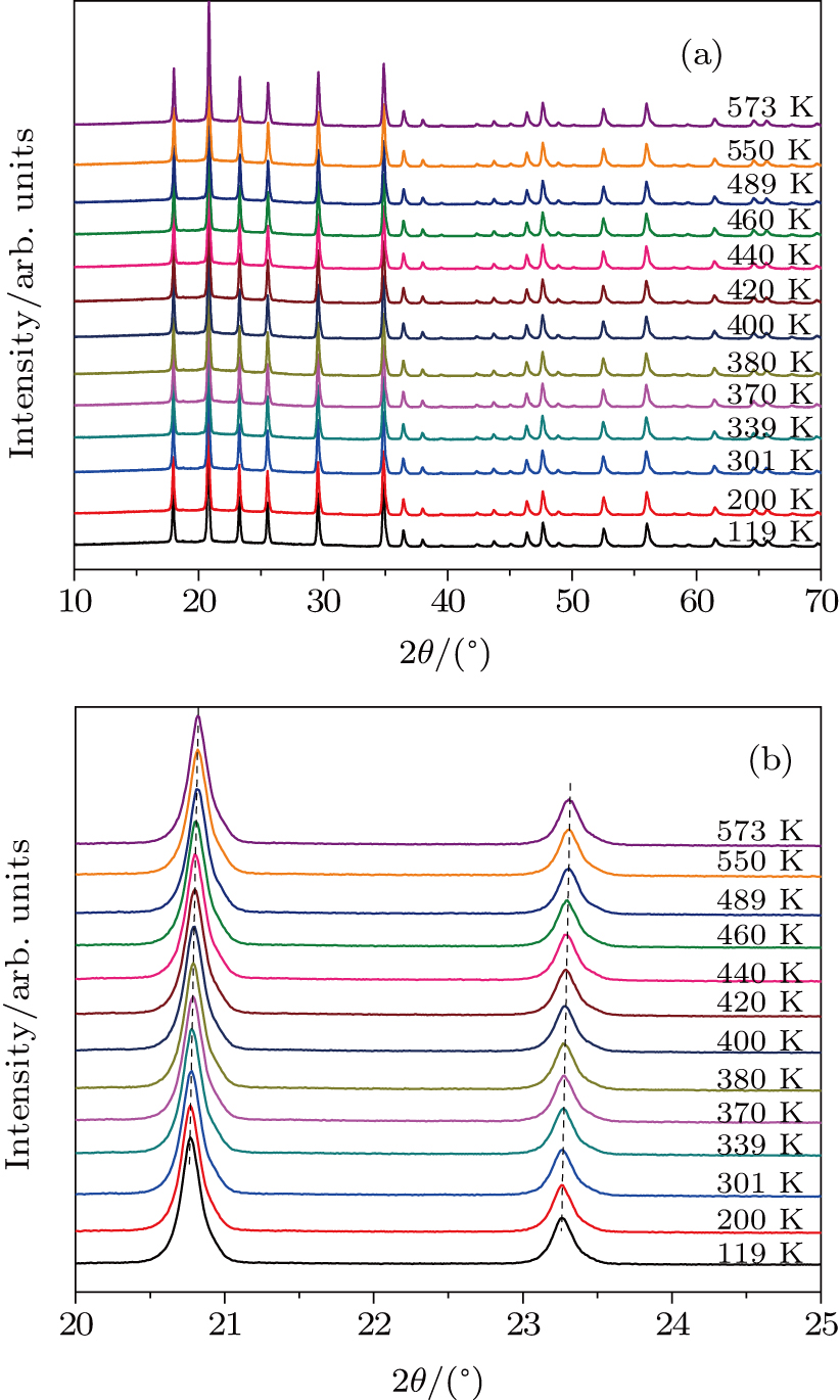
Figure 5 shows the temperature dependent XRD patterns of Zr0.5Hf0.5VPO7 in a temperature range from 119 K to 573 K. It is observed that the diffraction peaks of the XRD slightly shift to larger angles with temperature increasing (Fig. 5(b)). There are no obvious peaks appearing/disappearing in the XRD patterns in a temperature range from 119 K to 573 K. These results suggest that the samples keep cubic structures with temperature increasing. The cubic structures are found to be well consistent with space group .

Fig. 5. (color online) (a) XRD patterns of Zr0.5Hf0.5VPO7 at different temperatures. (b) Magnified part of patterns in panel (a).

Figure 6 exhibits the variation of the cell volume with temperature, which is calculated by Powder X. It can be clearly seen that there is no abrupt thermal expansion above 400 K. That is because Zr0.5Hf0.5VPO7 keeps a cubic structure in a temperature from 113 K to 673 K without phase transition. The CTE for volume is calculated to be K−1 in a temperature range of 400 K–573 K. This gives rise to a linear , confirming that Zr0.5Hf0.5VPO7 is intrinsically a ZTE material. This result is consistent with that from the Raman spectra, the 3 × 3 × 3 superstructure remarkably disappears for Zr0.5Hf0.5VPO7. The CTE for Zr0.5Hf0.5VPO7 is measured to be 0.59 × 10⁻⁶ K−1 in a temperature range of 364 K–673 K by dilatometry. The difference between CTEs measured by the dilatometer and XRD can relate to the microstructure effect which can add a small negative component to the intrinsic linear expansion coefficient.

Fig. 6. (color online) Variation of the cell volume of Zr0.5Hf0.5VPO7 with temperature.

The electronegativity of the cation and the M–O bond strength can be used to analyze the NTE of Zr0.5Hf0.5VPO7. The electronegativity value of P5+ (2.19) is larger than that of V5+ (1.63). The bridging oxygen connected with V5+ or P5+ gains less negative charges with the content of P5+ increasing, which reduces the effective negative charge due to the oxygen-oxygen repulsion force and makes the transition harder from superstructure to ideal crystal structure. The partial substitution of V in ZrV2O7 by P reduces the symmetry and influences the transverse thermal vibration of V–O–V. The bond angle of V (P)–O–V in ZrV2O7 is close to 180° because the transverse motion of bridge O in V–O–V and P–O–V is responsible for the negative thermal expansion of ZrV2O7.[1,17]

Dilatometric results are slightly different from the results from the variable-temperature XRD and variable-temperature Raman spectra. The microstructure effects might be responsible for the cause of such a difference. Raman spectra and XRD reveal the internal molecular structure and vibration mode of the unit cell. The Raman peak will change when the superstructure is disintegrated. The CTE from the dilatometer reflects the thermal expansion of bulk material, in which microcrack and poles are inevitable and may affect the measured results. The differences between intrinsic (XRD) and extrinsic (dilatometric) thermal expansion can be found in Zr1−xFexV2−xMoxO7,[7] ZrFeMo2VO12,[17] etc.

Figure 7 shows the SEM images of the sample. The microstructure of the grain morphology presents a spherical shape, which is closely arranged with the size of single short rod particle in a range from 2 μm to 6 μm. There are a few tiny gaps between rod particles. It should be noted that there are less pores in Zr0.5Hf0.5VPO7 than in other samples even under the same sintering conditions.

Fig. 7. SEM images of Zr0.5Hf0.5VPO7 synthesized by solid state reaction.
4. Conclusions

A solid solution of Zr0.5Hf0.5VPO7 with near-ZTE has successfully been synthesized by solid state reaction. Thermal dilatometry, XRD, and Raman spectra are used to investigate the compactness, the coefficient of thermal expansion, isotropy, and phase transition of Zr0.5Hf0.5VPO7. The results show that stable near-zero thermal expansion of the prepared material goes over a wide temperature range. The CTE for Zr0.5Hf0.5VPO7 is measured to be 0.59 × 10⁻⁶ K−1 in a temperature range of 310 K–673 K by dilatometry while it is calculated to be 0.259 × 10⁻⁶ K−1 (400 K–573 K) by XRD. The results from the dilatometer are slightly different from those from the variable-temperature XRD. The differences between the two methods are attributed to the effects of microstructure.

Reference
[1] Yuan B H Liu X S Mao Y C Wang J Q Guo J Cheng Y G Song W B Liang E J Chao M J 2016 Mater. Chem. Phys. 170 162
[2] Dai Y J Huang R J Wang W Li L F Sun Z H 2017 Ceram. Int. 43 1608
[3] Ding L Wang C Na Y Y Chu L H Yan J 2011 Scr. Mater. 65 687
[4] Yan J Sun Y Wang C Chu L H Shi Z X Deng S H Shi K W Lu H Q 2014 Scr. Mater. 84-85 19
[5] Yan X Cheng X N Yang X B Zhang C H 2007 Key Eng. Mater. 353-358 1235
[6] Yang X B Cheng X N Yan X H Yang J Fu T B Qiu J 2007 Compos. Sci. Technol. 1167-1171 67
[7] Yang X B Xu J Li H J Cheng X N Yan X H 2007 J. Am. Ceram. Soc. 90 1953
[8] Romao C P Perras F A Wernerzwanziger U Lussier J A Miller K J 2015 Chem. Mater. 27 2633
[9] Liang E J Liang Y Zhao Y Liu J Jiang Y J 2008 J. Phys. Chem. 112 12582
[10] Yuan B H Liu X S Mao Y C Wang J Q Guo J Cheng Y G Song W B Liang E J Chao M J 2016 Mater. Chem. Phys. 170 162
[11] Yanase I Kojima T Kobayashi H 2011 Solid State Commun. 151 595
[12] Mittal R Chaplot S L 2008 Phys. Rev. 78 2599
[13] Yuan H L Yuan B H Li F Liang E J 2012 Acta Phys. Sin. 61 226502 in Chinese
[14] Song W B Liang E J Liu X S Li Z Y Yuan B H Wang J Q 2013 Chin. Phys. Lett. 30 126502
[15] Wu M M Peng J Zu Y Liu Y D Hu Z B Liu Y T Chen D F 2012 Chin. Phys. 21 116102
[16] Li Q J Yuan B H Song W B Liang E J Yuan B 2012 Chin. Phys. 21 046501
[17] Hisashige T Yamaguchi T Tsuji T Yamamura Y 2006 J. Ceram. Soc. Jpn. 114 607
[18] Evans J S O Hanson J C Sleight A W 1998 Acta Crystallogr. 54 705
[19] Xiang H M Feng Z H Zhou Y C 2014 J. Eur. Ceram. Soc. 34 1809
[20] Jiang X R Molokeev M S Gong P F Yang Y Wang W Wang S H et al. 2016 Adv. Mater. 28 7936
[21] Chen D X Yuan B H Cheng Y G Ge X H Jia Y Liang E J et al. 2016 Phys. Lett. 380 4070
[22] Marinkovic B A Jardim P M Ari M Avillez R R D Rizzo F Ferreira F F 2008 Phys. Stat. Sol. 245 2514
[23] Hemamala U L C El F Muthu D V S Krogh A M Andersen Carlson S Ouyang L Kruger M B 2007 Solid State Commun. 141 680