A computational study of the chemokine receptor CXCR1 bound with interleukin-8
Wang Yang1, Severin Lupala Cecylia1, Wang Ting2, Li Xuanxuan1, 3, Yun Ji-Hye4, Park Jae-hyun4, Jin Zeyu4, Lee Weontae4, Tan Leihan1, 5, Liu Haiguang1, †
       

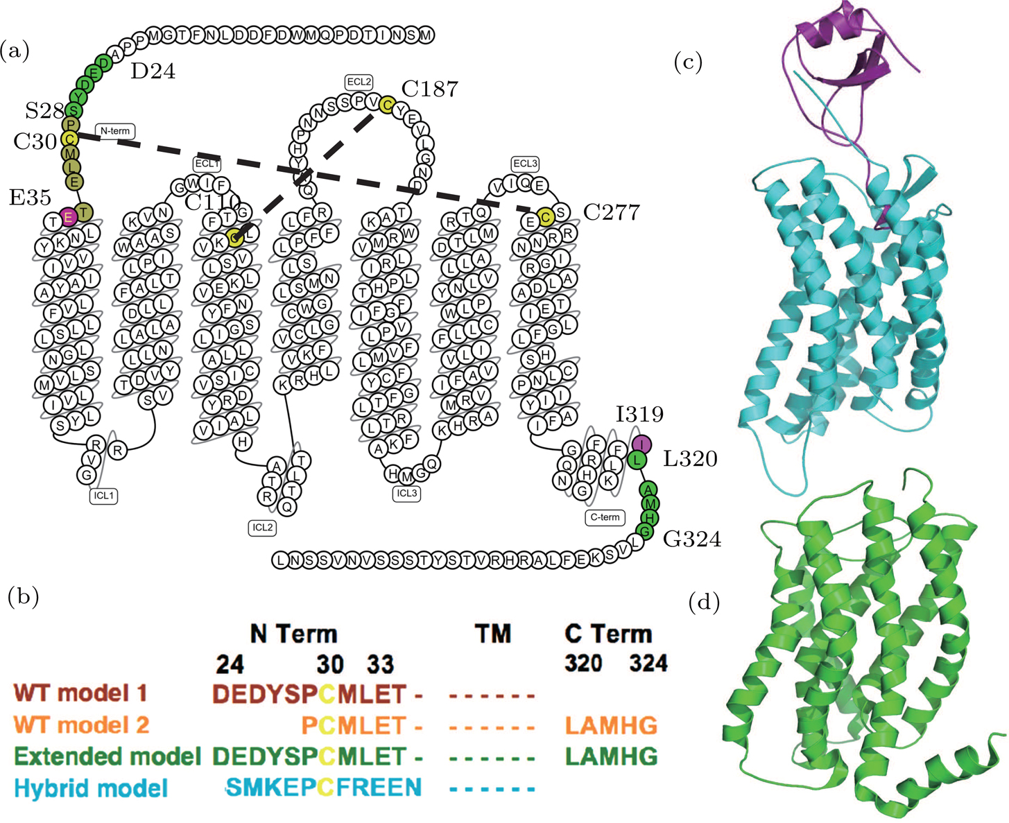
(color online) The CXCR1-IL8 systems. (a) The sequence and secondary structure of CXCR1. The snake map is drawn from GPCR db.[32] The residues (C30, C110, C187, and C277) that form disulfide bonds are colored in yellow. The sequence between two purple residues E35 and I319 forms the main body of the CXCR1 receptor. (b) Sequence differences of four constructed models. The four models are colored in brown (WT model 1), orange (WT model 2), green (extended model), and cyan (hybrid model), respectively. (c) The crystal structure of CXCR4-vMIP-II binding complex (PDB code: 4RWS). (d) The NMR structure of CXCR1 (PDB code: 2LNL).