Areal density and spatial resolution of high energy electron radiography
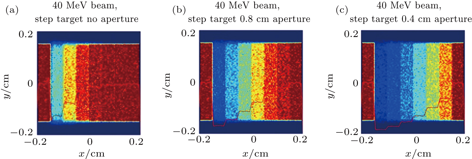
(color online) Simulated images of the step target with (a) no aperture, (b) 8 mm size aperture, and (c) 4 mm size aperture at the Fourier plane in the
Areal density and spatial resolution of high energy electron radiography |
|
(color online) Simulated images of the step target with (a) no aperture, (b) 8 mm size aperture, and (c) 4 mm size aperture at the Fourier plane in the |
| |