Entangled-photons generation with quantum dots
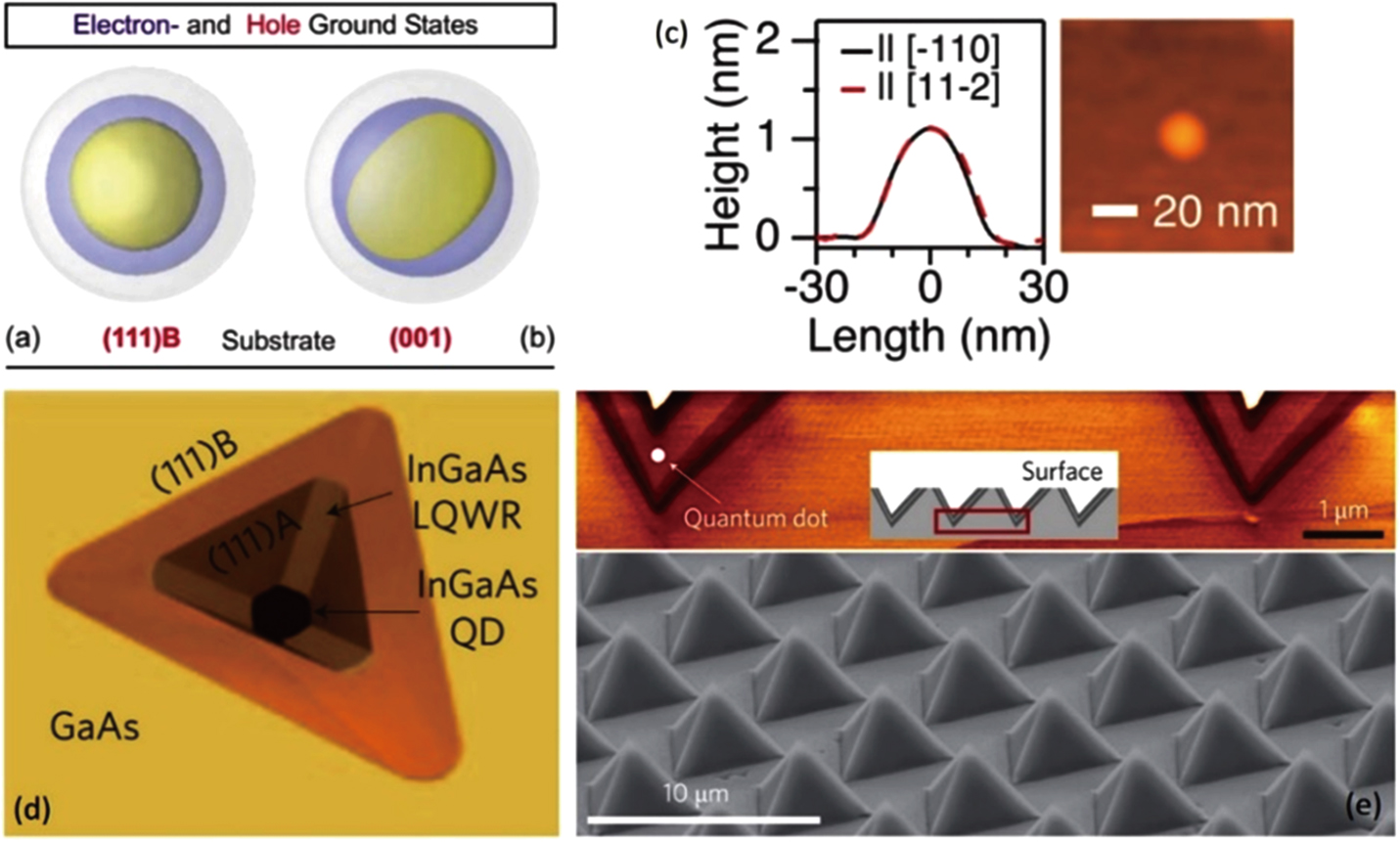
(color online) (a) and (b) Orientation of electron (blue) and hole (yellow) wave functions for a lens-shaped QD on two substrates with different orientations.[

Entangled-photons generation with quantum dots |
|
(color online) (a) and (b) Orientation of electron (blue) and hole (yellow) wave functions for a lens-shaped QD on two substrates with different orientations.[ |
 |