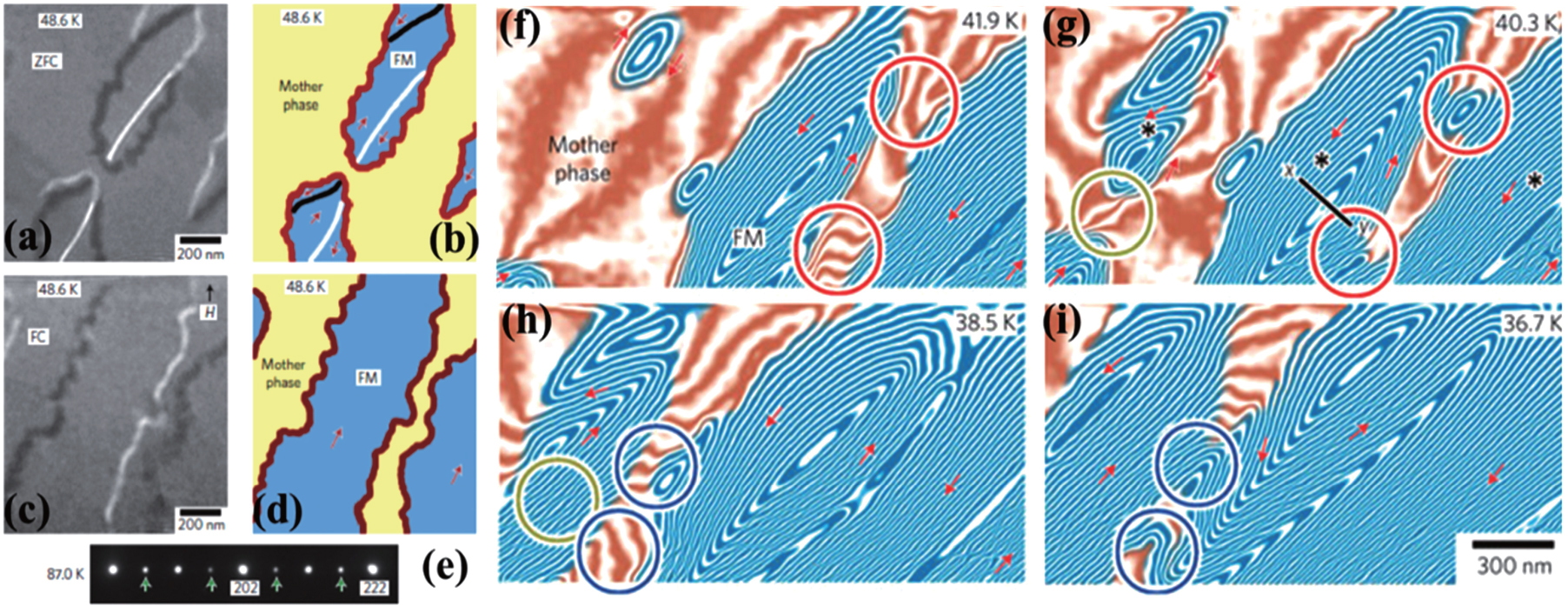
Microscopic observation of phase boundaries and the coalescence of FM domains using cryogenic Lorentz microscope and electron holography. Lorentz microscope images of multiple domains and a single domain formed in ZFC (a) and FC (c). (b), (d) The corresponding schematics of the boundary structures in panels (a) and (c). The red lines in panels (b) and (d) depict the phase boundaries, and the black and white lines in panel (b) represent the magnetic domain walls within multiple FM domains. The red arrows in panels (b) and (d) indicate the magnetization vectors determined by the holographic analysis. The black arrow in panel (c) represents the direction of the applied magnetic field H in the FC measurement. The Lorentz microscope images were taken in the under-focus condition. (e) Electron diffraction pattern observed in the mother phase at 87 K. The green arrows indicate superlattice reflections, which indicate the presence of the CO portions within the mother phase. (f)–(i) ZFC observations of the coalescence of FM domains distributed independently in the mother phase. Regions having blue lines are in the FM phase and those having sepia lines are in the mother phase. Contour lines and the red arrows represent the in-plane component and the direction of the magnetic flux, respectively. Domain connection occurs preferentially in areas (indicated by circles) where the leakage magnetic field is relatively strong. The phase information has been amplified by a factor of four. The red circles, green circles, and blue circles represent domain coalescence that was observed in cooling from 41.9 K to 40.3 K, from 40.3 K to 38.5 K, and from 38.5 K to 36.7 K, respectively. Reprinted with permission from Ref. [32]. Copyright 2009, Nature Publishing Group. |

