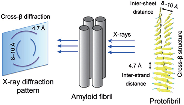
A diagram showing the x-ray diffraction pattern and cross-βstructure feature of fibrils. The characteristic cross-βdiffraction pattern is generated when x-rays are directed on amyloid fibrils, which consist of multiple protofibrils. The diffuse reflection at 4.7 Å spacing along the meridian (vertical) shows extended protein chains running approximately perpendicular to the fibril axis and spaced 4.7 Å apart. The increasingly diffuse reflection at 8–10 Å spacing along the equator (horizontal) shows that the extended chains are organized into sheets spaced 8–10 Å apart. The cross-βstructure can be seen from the cartoon representation of a protofibril. |