A review of the growth and structures of silicene on Ag (111)
Wu Ke-Hui†a), b) 
A review of the growth and structures of silicene on Ag (111) |
|
Wu Ke-Hui†
|
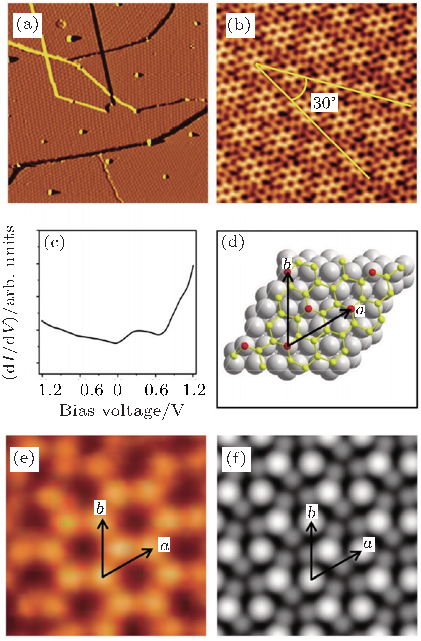
| (a) A derivative STM image (200 nm×200 nm, V tip = 1.43 V) of a surface fully covered by the phase. (b) High-resolution STM image (15 nm×15 nm, V tip = −1.0 V) showing the atomic structure. The bright areas exhibit complete honeycomb rings with a period of 1.0 nm, while other areas are disordered. (c) d I /d V spectra shows a peak at 0.3 V and a shoulder at 0.9 V are observed. (d) Calculated model of superstructure of silicene. The gray, yellow, and red balls represent the silver, lower silicon, and higher silicon atoms, respectively. (e) and (f) Experimental and simulated STM images (1.0 eV above Fermi energy) showing the similar structure features and unit cell of lattice (adapted from Ref. [ 14 ]). |
| |