Experimental research on the feature of an x-ray Talbot–Lau interferometer versus tube accelerating voltage

Experimental research on the feature of an x-ray Talbot–Lau interferometer versus tube accelerating voltage |
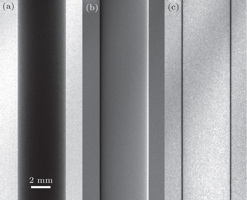
| The x-ray imaging results of the PMMA cylinder obtained at an accelerating voltage of 35 kV, showing (a) conventional absorption image, (b) refraction signal, and (c) normalized visibility image. All the images are windowed for optimized appearance with a linear gray scale. |
 |