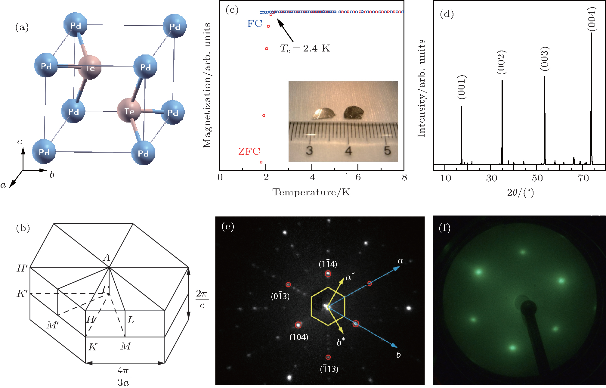
Electronic structure of transition metal dichalcogenides PdTe2 and Cu0.05PdTe2 superconductors obtained by angle-resolved photoemission spectroscopy*
Liu Yana), Zhao Jian-Zhoua), Yu Lia), Lin Cheng-Tianb), Hu Chenga), Liu De-Faa), Peng Ying-Yinga), Xie Zhuo-Jina), He Jun-Fenga), Chen Chao-Yua), Feng Yaa), Yi He-Miana), Liu Xua), Zhao Lina), He Shao-Longa), Liu Guo-Donga), Dong Xiao-Lia), Zhang Juna), Chen Chuang-Tianc), Xu Zu-Yanc), Weng Hong-Minga), Dai Xia), Fang Zhonga), Zhou Xing-Jianga),d)†
       
Crystal structure and Brillouin zone of PdTe2 and sample characterization of Cu0.05PdTe2 single crystal. (a) Crystal structure of PdTe2 with a polymeric CdI2-type structure (space group: P 3 m 1). (b) The corresponding hexagonal prism Brillouin zone of PdTe2. (c) Magnetization measurement of Cu0.05PdTe2 single crystal with a magnetic field of 1 Oe. FC (blue circles) refers to the field-cooled measurement, while ZFC (red circles) refers to the zero-field-cooled measurement. The bottom-right inset shows a photograph of the plate-like Cu0.05PdTe2 single crystal with a shining surface and a dimension of 5 mm×3 mm×0.2 mm. (d) The x-ray diffraction pattern of a naturally cleaved surface of Cu0.05PdTe2 single crystal, showing that it is the basal plane. (e) Laue diffraction pattern of the Cu0.05PdTe2 single crystal at room temperature. The six red circles mark the six spots we used for indexing search by Laue simulation. The blue axes are the determined lattice vectors a and b in the real space from the Laue indexing. The yellow axes are the determined reciprocal lattice vectors a * and b *. The yellow hexagon is the first Brillouin zone determined by the reciprocal lattice vectors. (f) The low-energy electron diffraction pattern of a freshly cleaved Cu0.05PdTe2 surface taken with an electron energy of 70 eV at 25 K.