Giant magnetic moment at open ends of multiwalled carbon nanotubes

Giant magnetic moment at open ends of multiwalled carbon nanotubes |
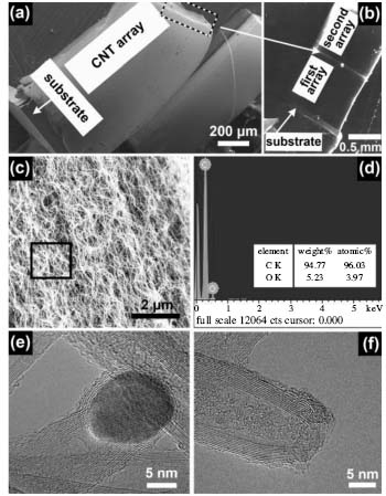
| Fig. 2. a SEM image showing CNT arrays grown from the substrate. The iron catalytic nanoparticles are shown to be at the tips of the CNTs because the tips of the 1 st CNT array can be used as the substrate to grow the 2 nd CNT array, as shown in panel b. c and d SEM image of the CNTs in the middle part of the array and the corresponding EDX results from the marked area in panel c, indicating there is no iron found in the central part of a CNT array. e HRTEM high resolution transmission electron microscope image showing that at the tip of a CNT, there is an iron catalytic nanoparticle. f Typical HRTEM image showing that the end of CNT is open after the CNT is cut. |
 |