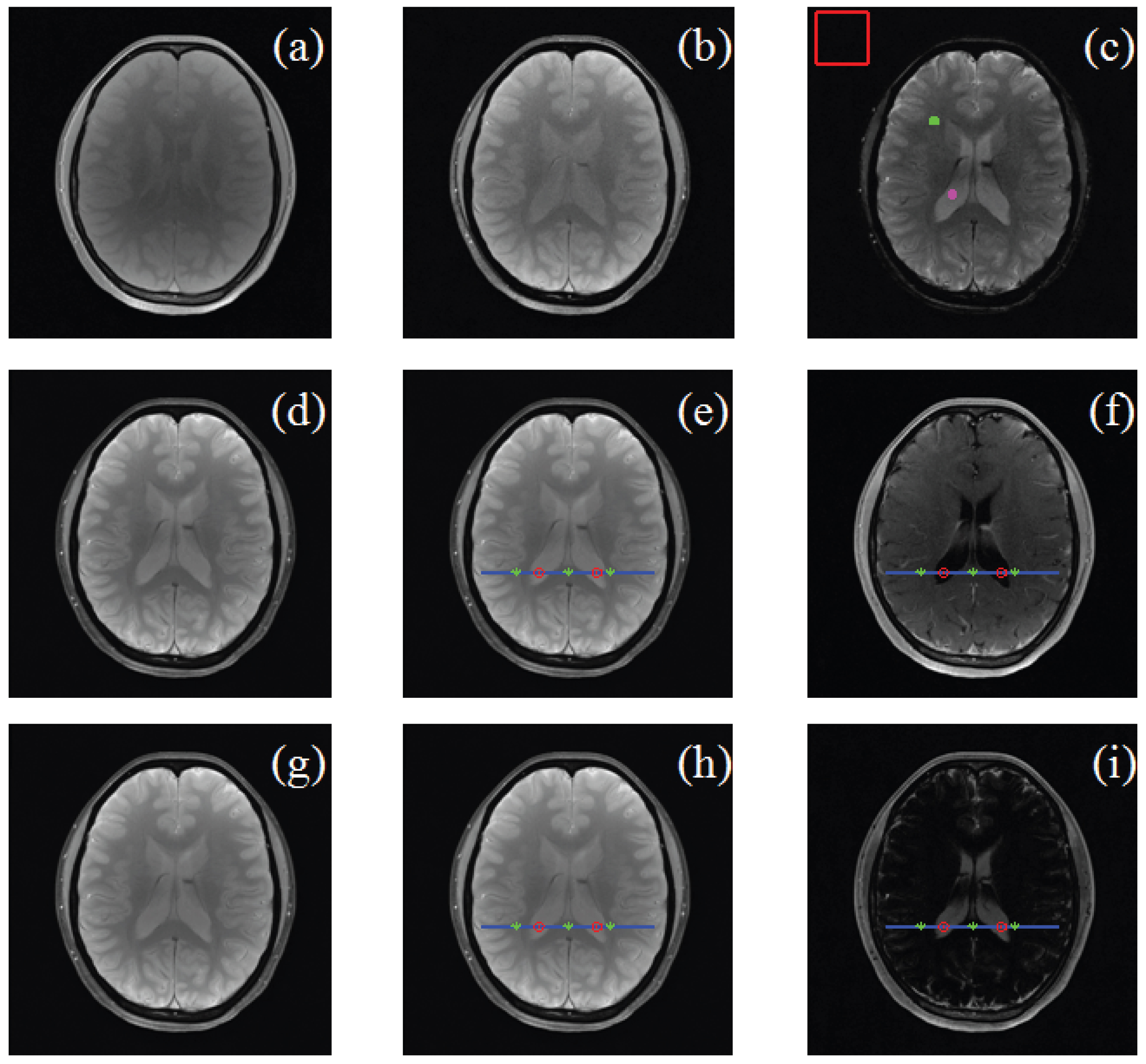
Linear-fitting-based similarity coefficient map for tissue dissimilarity analysis in T2*-w magnetic resonance imaging
Yu Shao-Dea),b), Wu Shi-Bina),b), Wang Hao-Yuc), Wei Xin-Huad), Chen Xind), Pan Wan-Longe), Hu Jianif), Xie Yao-Qin†a)
       
Perceived visual image quality analyses. The third image series of the first volunteer and its derived maps are represented. Panels (a), (b), and (c) are -w magnetic resonance images with ascending TE value (2.61 ms, 12.51 ms, and 35.61 ms), panels (d), (e), and (f), and panels (g), (h), and (i) are SCM, RMM, and RM maps from tissue of interest (CSF and then WM) as the reference tissue, respectively. Pixels on the blue lines are for further intra- and inter-tissue pixel intensity investigation.