Enhancement of photoacoustic tomography in the tissue with speed-of-sound variance using ultrasound computed tomography
Cheng Ren-Xiang, Tao Chao , Liu Xiao-Jun
, Liu Xiao-Jun 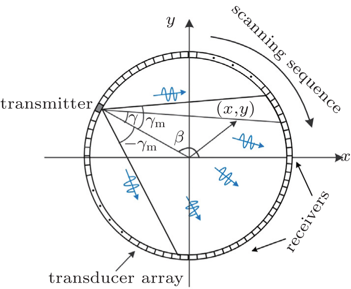
Enhancement of photoacoustic tomography in the tissue with speed-of-sound variance using ultrasound computed tomography |
|
Cheng Ren-Xiang, Tao Chao
, Liu Xiao-Jun |
| Schematic of scanning geometry. A US transducer element at angular position β is used as the transmitter to launch US pulses. The other elements on the opposite side of the transmitter are used as the receivers to detect the US pulses. The process is repeated to make the fan-beam (light gray region) rotate 360 degrees around the tissue. |
| |