Flexible reduced field of view magnetic resonance imaging based on single-shot spatiotemporally encoded technique
Li Jinga) , Cai Cong-Bob) , Chen Lina) , Chen Yingc) , Qu Xiao-Boa) , Cai Shu-Hui†a) 
Flexible reduced field of view magnetic resonance imaging based on single-shot spatiotemporally encoded technique |
|
Li Jing
|
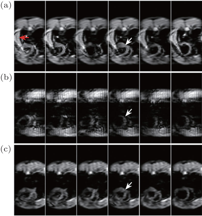
| In vivo rat cardiac images. (a) EPI images. (b) SPEN images ( P = 1). (c) The rFOV images obtained with the flexible rFOV imaging method. Spatiotemporal encoding is conducted along the horizontal direction, slice thickness = 2 mm, L x = 50 mm (vertical), sampling data matrix size = 32 × 64 (horizontal × vertical), image matrix size after SR reconstruction = 32 × 128, sequence execution time = 30.4 ms, Δ O Hz = 64 kHz, T exc = 4 ms, T acq = 13.9 ms, P = 0.5, L y = 44 mm (horizontal), L yd = 22 mm, the interval between two successive frames = 8 s, R = 16 kHz/ms, and acquisition bandwidth along readout dimension = 250 kHz. |
| |