Flexible reduced field of view magnetic resonance imaging based on single-shot spatiotemporally encoded technique
Li Jinga) , Cai Cong-Bob) , Chen Lina) , Chen Yingc) , Qu Xiao-Boa) , Cai Shu-Hui†a) 
Flexible reduced field of view magnetic resonance imaging based on single-shot spatiotemporally encoded technique |
|
Li Jing
|
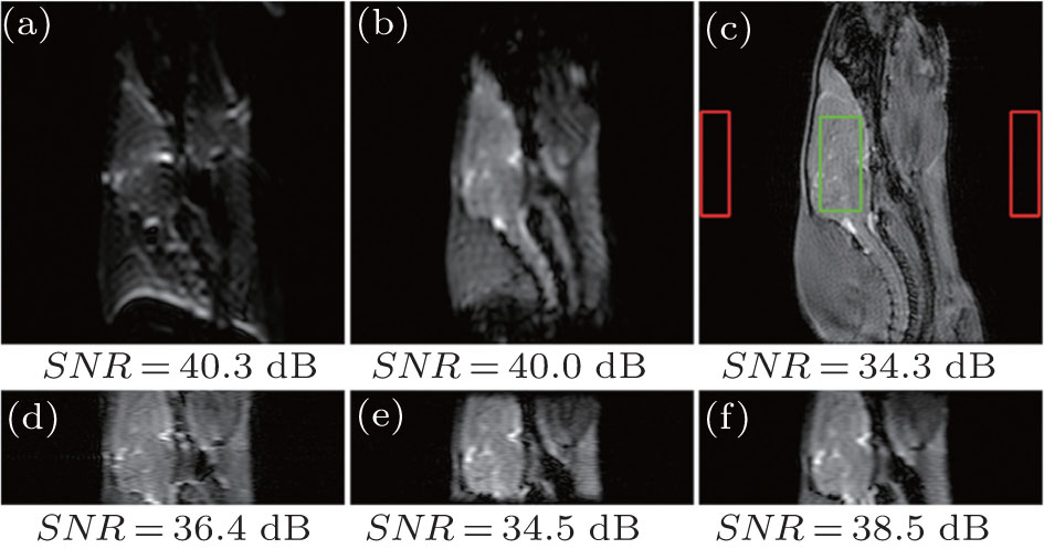
| Imaging results of a sagittal plane of an in vivo rat head. Slice thickness = 2 mm, imaging matrix size = 256 × 256, L x = 60 mm (horizontal), L y = 60 mm [(a)–(c)], 20 mm [(d)–(f)] (vertical). (a) and (d) EPI images (sampling data matrix size = 64 × 64, G acq = 0.0196 T/m (a), 0.0367 T/m (d), sequence execution time = 47.6 ms). (b) and (e) SPEN images ( P = 1, G acq = 0.0367 T/m (b), 0.0587 T/m (e)). (c) Reference multi-scan gradient echo image (sampling data matrix size = 128 × 128, repetition time ( TR ) = 100 ms, dummy scans = 16, sequence execution time = 14.4 s). (f) SPEN image obtained with the flexible rFOV imaging method ( P = 0.625, G acq = 0.0311 T/m). Common parameters for SPEN imaging: T exc = 3 ms, Δ O Hz = 64 kHz, sampling data matrix size = 64 × 64, R = 21.3 kHz/ms, sequence execution time = 49.1 ms, and acquisition bandwidth along readout dimension = 250 kHz. |
| |