


Tunable Fano resonances and plasmonic hybridization of gold triangle–rod dimer nanostructure
[Huang Meng1, †,  , Chen Dong2, Zhang Li2, Zhou Jun2]
, Chen Dong2, Zhang Li2, Zhou Jun2]
|
|


† Corresponding author. E-mail:
Project supported by the National Natural Science Foundation of China (Grant No. 61275153) and the Natural Science Foundation of Zhejiang Provice, China (Grant No. LY12A04002).
A gold dimer structure consisting of a notched triangle nanoslice and a rectangle nanorod is proposed to produce distinct Fano resonance. Owing to the coupling between the dipole plasmon mode of the nanorod and the dipole or quadrupole plasmon mode of the nanoslice, the extinction spectrum with a deep Fano dip is formed and can be well fitted by the Fano interference model for different geometry parameters. In addition, Fano resonance of the gold dimer nanostructure also intensely depends on the polarization direction of incident light. Moreover, Fano resonance of the triangle–rod trimer is also analyzed by adding another nanorod into the former dimer and exhibits the splitting of plasmonic resonant peak in high order coupling modes. The plasmonic hybridizations in these nanostructures have been analyzed for revealing the physical origin of the Fano resonance.
As is well known, the optical properties of noble metallic nanostructures are dominated by the excited localized surface plasmon resonance (LSPR) when the frequency of the incident electromagnetic wave is virtually identical to the vibration frequency of collective electron in the nanostructure.[1] The frequency, strength, and quality of the LSPR depend on the size, geometry, and composition of an individual metallic nanostructure, and also relate to the refractive index of the local environment.[2–4] In particular, the LSPR characteristic of a metal nanoparticle is intensively sensitive to the presence of other nearby metal nanoparticles. When two LSPR bands are overlapped and coupled through near-field interactions, splitting of the modal energies can occur and the bonding and antibonding modes are formed according to the plasmon hybridization theory.[5] Thence, for obtaining tunable richer plasmonic features, not only metallic nanostructures with different shapes such as spheres,[6] rods,[7] rings,[8,9] disks,[10] cube,[11] and shells,[12–14] but also the complex nanostructures with two or more elementary metallic shapes have been theoretically and experimentally investigated.[15–18] As a typical example, Fano resonance of a metallic nanostructure with complex or asymmetric geometry has become a research focus.[19–21] In fact, Fano resonance is caused by the interactions of a broad “bright” plasmon mode and a relatively narrow “dark” plasmon mode in metallic nanostructure and exhibits an asymmetric extinction line shape, and the feature of Fano resonance is highly sensitive to the geometric parameters of nanostructure, the angle of incidence light and the dielectric permittivity of environment,[22–24] so that a small perturbation can induce the dramatic shift of resonance lineshape. It is the above property to render many promising advantages of Fano resonance in actual applications such as chemical or biological sensors,[25] electromagnetically induced transparency (EIT),[26] switching,[27] plasmonic nanolasing,[28] and slow light.[29]
In recent years, besides the simple dimer nanostructures including sphere–sphere dimer,[30] sphere–rod dimer,[31] rod–rod dimer,[32] rod–ring disks dimer,[33–35] the plasmonic properties of other symmetry-breaking dimers[36] and the heterodimers composed of silver, gold, and copper nanoparticles have been widely studied because of their outstanding advantages of tunable plasmon resonance which intensely depend on polarization angles of incident light, gap of interparticle separations, especially, their multipolar Fano features are easily excited by hybridizing of a bonding quadrupole–quadrupole mode and a dipole–quadrupole mode.[37] For example, the plasmonic responses (including Fano resonances) of heterodimers with an Au nanorod and a small Au nanosphere, are remarkably sensitive to the nanosphere position on the nanorod, the gap distance, and the nanocrystal dimensions. As the nanosphere is moved around the nanorod, the rotational symmetry of the Au nanorod–nanosphere heterodimer is broken, which causes the coupling of plasmon modes and the varying of Fano interference.[31] The Fano resonance of T-shaped nanorod dimer or angle–resolved nanorod dimer can induce two splitting hybridized bonding and antibonding resonance modes which are affected by the geometry parameters and the incident polarization.[32] Yang et al. reported that the Fano resonance in gold ring–rod nanocavities with more than one nanorod, i.e., the plasmon coupling in the theta-shaped ring–rod cavity exhibits a flexible tunability.[34] Meanwhile, the narrow and deep Fano dip in LSPR spectrum can be achieved in the nanostructures with a rod and concentric square ring-disk.[35] The extinction properties of a plasmonic nanocavity consisting of a regular triangle embedded in a split ring are also sensitive to the polarization of the incident light, which results in strong Fano resonances.[36] Therefore, it is interesting to pay attention on the Fano resonances of the dimer with special shaped metal nanostructure.
In this paper, we design a gold dimer consisting of a notched triangle nanoslice and a cuboid nanorod to produce strong Fano resonances. The Fano resonances of gold dimer are formed by the plasmonic coupling between the “bright” dipole mode of the nanorod and “bright” dipole or the “dark” quadrupole modes of nanoslice, respectively. By using the Fano interference model and finite element method (FEM), the effects of the structure parameters and the incident polarization on the Fano resonances are analyzed for understanding the underlying physics of the Fano resonances in this dimer. In addition, compared with the works described above, the split of Fano resonance profile can be easily formed by simply extending the dimer to a trimer with another cuboid nanorod and used as a multi-wavelength high sensitive sensor in the near-infrared region.
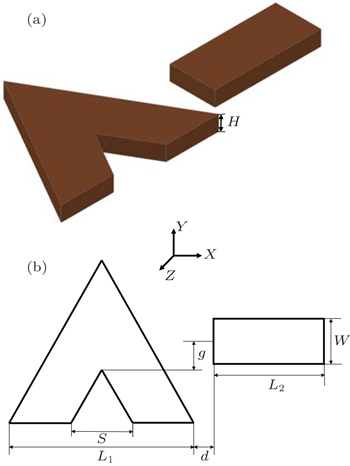
The schematic graph of the proposed gold triangle–rod dimer nanostructure is plotted in Fig.
 | Fig. 1. (a) Model configuration of the proposed dimer and (b) geometric parameters of the notched triangle nanoslice and the rectangle nanorod: L1 = 130 nm, L2 = 120 nm, W = 60 nm, H = 30 nm, S = L1/3 = 43.3 nm. |
By using the scattering field formulation, the optical properties of the proposed gold triangle–rod nanostructure can be numerically studied in the frequency domain and be well expressed by the scattering cross-section (Cscat), the absorption cross-section (Cabs), and the extinction cross-section (Cext), respectively. That is that Cscat is obtained by integrating the normalized electric field around a far-field transform boundary enclosing the dimer. Similarly, Cabs is obtained by integrating the time-average resistive heating (Uav). Here, Cscat and Cabs are expressed as follows:[40]

In fact, the extinction cross-section Cext of the system is defined as the total radiation flux of scattering and absorption transmitting through the system. We have

In our research, the commercial software package COMSOL Multiphysics 4.3 incorporated RF module, which contains electromagnetic scattering formula code based on the three-dimensional finite element method (3D-FEM), was used to calculate the extinction cross section, near-field intensity distributions, and charge distributions. As we know, FEM is an effective research method of the near-field optics and suitable to analyze the optical properties of metallic nanostructures. It is said that the electromagnetic problems of complex nanostructures can be effectively handled by solving Maxwell’s equations on an appropriate discretized spatial grid.[41,42] In our numerical simulations, the perfectly matched layers were used in the propagation direction to eliminate the nonphysical reflections at the domain boundaries, and the triangle–rod dimer nanostructure was meshed with tetrahedral elements and a local mesh refinement was used in and around the dimer.
For the different polarizations of incident light, the extinction cross-section spectra of the individual notched triangle nanoslice and nanorod are shown in Fig.
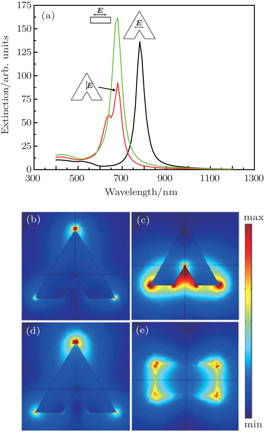 | Fig. 2. (a) Extinction spectra of the individual gold nanorod and a notched triangle nanoslice. Here, |
Factually, for the case of the polarization of incident light along the horizontal direction, the dipole plasmon modes in the nanoslice and the nanorod are excited into bright modes with lower energies. The quadrupole plasmon mode in the nanoslice cannot be excited as a dark mode in the far field by the horizontal polarization light, but it can be excited effectively by the near-field plasmon coupling between the dipole mode of the nanorod and the quadrupole of the nanoslice. By bringing the nanorod and nanoslice close to each other, the bright dipole mode of nanorod and the dark quadrupole mode of the nanoslice can overlap and destructively interfere at the resonant wavelength to form an Fano dip in the extinction spectrum.
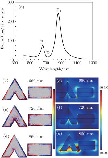
On the other hand, it is important to discuss the lineshape feature of Fano resonance and the electric field distributions of the dimer nanostructure. Figure
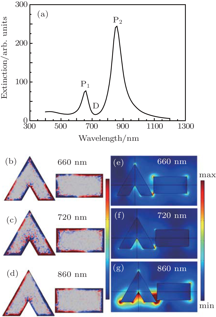 | Fig. 3. (a) Extinction spectrum of the triangle–rod dimer nanostructure with a geometric center separation g = 0 nm and a gap distance d = 2 nm; (b)–(d) The charge distributions at the Fano peak P1, Fano dip D, Fano peak P2, respectively; (e)–(g) The electric field distributions at Fano peak P1, Fano dip D, Fano peak P2, respectively. |
Furthermore, the Fano resonance characteristic of the proposed dimer can be tuned by adjusting the geometry of the triangle–rod nanostructure. Figure
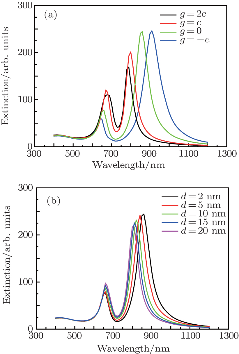 | Fig. 4. (a) Extinction spectra of the triangle–rod dimer nanostructure with varying the center offset g for the fixed gap distance d = 2 nm; (b) extinction spectra of the triangle–rod dimer nanostructure with varying the gap distance d and the fixed center offset g = 0 nm. The constant c equals to the vertical distance between the center and the side of notched triangle nanoslice. |
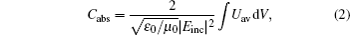
The Fano resonance characteristic of the proposed dimer is also dependent on the polarization direction of incident light. As shown in Fig.
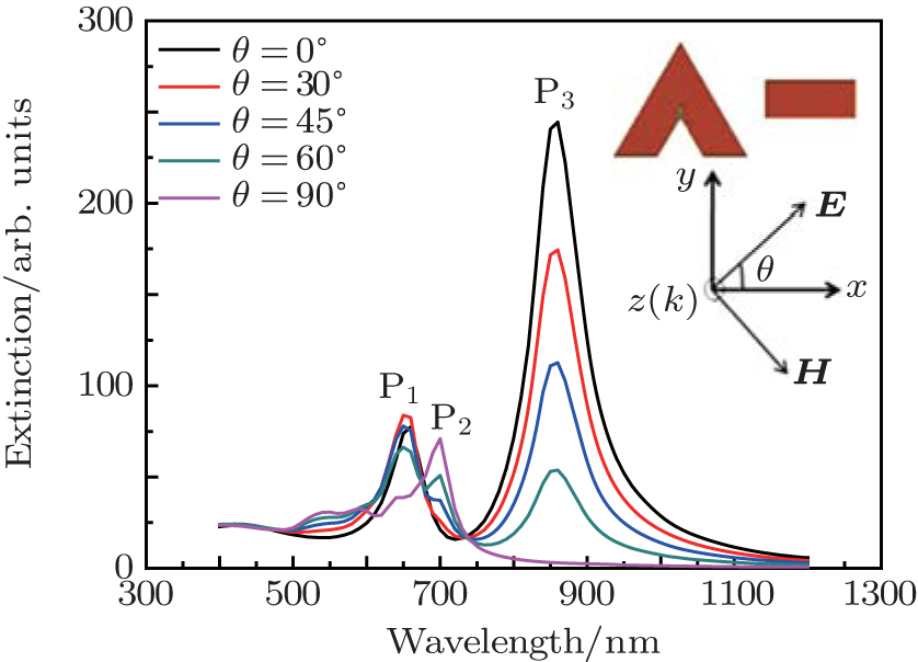 | Fig. 5. The incident polarization effects on the extinction spectra of the triangle–rod dimer nanostructure (g = 0 nm, d = 2 nm). The inset shows incident polarizations. |
It is more interesting that the extinction spectrum of the triangle–rod trimer nanostructure demonstrates the perfect profile of Fano resonance. The triangle–rod trimer consists of two nanorods and a notched triangle nanoslice and is drawn in Fig.
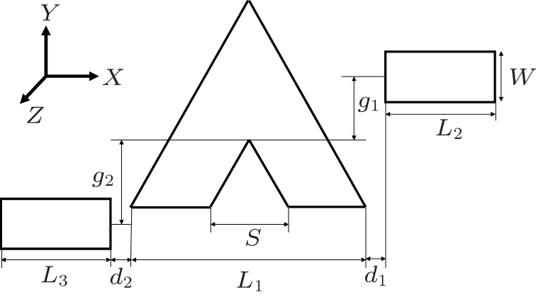 | Fig. 6. The configuration of an Au triangle–rod trimer with geometric parameters: L1 = 130 nm, L2 = 120 nm, L3 = 120 nm, W = 60 nm, H = 30 nm, and S = L1/3 = 43.3 nm. |
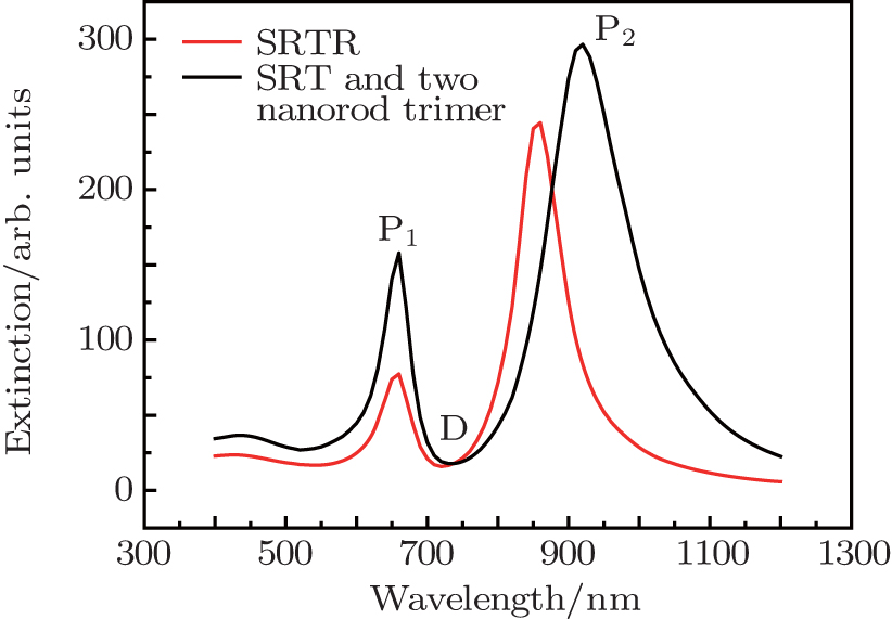 | Fig. 7. The extinction spectrum of the proposed dimer and trimer for d1 = d2 = 2 nm and g1 = g2 = 0. |
 | Fig. 8. The extinction spectra of the proposed trimer (a) for varying position of right nanorod g1 at d1 = d2 = 2 nm and g2 = 0 and (b) for varying length of left nanorod L3 at d1 = d2 = 2 nm and g1 = g2 = 0. The electric field distributions at resonant peaks (c) P1, (d) P2, and (e) P3, for L3 = 170 nm, respectively. All the other structure parameters of the trimer are the same as those in Fig. |
A simple plasmonic gold triangle–rod dimer nanostructure consisting of a regular triangle nanoslice with a triangle split and a nanorod is proposed and its strong Fano resonance has been simulated by FEM. From the viewpoint of plasmon hybridization theory, the Fano resonance of the proposed nanosystem is the interference result between the “bright” dipole mode of the rod and the “bright” dipole and the “dark” quadrupole modes in the hybrid nanostructure, that is, the interaction among the plasmon modes leads to their coupling mode energy to be split into a high-energy anti-bonding mode and a low-energy bonding mode. The simulation results show that the extinction spectra of the dimer intensively depend on its geometric parameters. In particular, the resonant peak of low-energy bonding mode red-shifts but the resonant peak of high-energy anti-bonding mode blue-shifts with the decrease of the center offset or the coupling distance between the nanoslice and the nanorod. In addition, the intensities of Fano resonant peaks of the dimer are decreased with the increase of the polarization angle of incident light. Based on the dimer structure, a more perfect Fano resonance can be derived by a gold triangle–rod trimer nanosystem which simply adds another nanorod into the original dimer structure. The electric field distributions of the dimer and the trimer demonstrate an almost identical trend with varying of structure parameters, which reveals the same physics origin from the plasmonic hybridization or interference in the nano-assemblies. Our results are helpful to design a simple nanostructure with excellent performance of Fano resonance in the chemical and bio-sensing application.
| 1 | |
| 2 | |
| 3 | |
| 4 | |
| 5 | |
| 6 | |
| 7 | |
| 8 | |
| 9 | |
| 10 | |
| 11 | |
| 12 | |
| 13 | |
| 14 | |
| 15 | |
| 16 | |
| 17 | |
| 18 | |
| 19 | |
| 20 | |
| 21 | |
| 22 | |
| 23 | |
| 24 | |
| 25 | |
| 26 | |
| 27 | |
| 28 | |
| 29 | |
| 30 | |
| 31 | |
| 32 | |
| 33 | |
| 34 | |
| 35 | |
| 36 | |
| 37 | |
| 38 | |
| 39 | |
| 40 | |
| 41 | |
| 42 |