

Fluorescence enhancement of radix angelica dahurica by binding to single silver sphere*
[Wang Qing-Rua), b)†  , Shi Qiang
a), b) , Li Shu-Honga), b) , Wang Wen-Juna), b) ]
, Shi Qiang|
|


†Corresponding author. E-mail: wangqingru@lcu.edu.cn
*Project supported by the National Natural Science Foundation of China (Grant Nos. 61405085 and 61275147), the Research Fund for the Doctoral Program of Liaocheng University, China, the Key Project of Science and Technology of Shandong Province of China (Grant No. 2010GGX10127), and the Shandong Province Natural Science Foundation of China (Grant Nos. ZR2013EML006 and ZR2012AL11).
We present a theoretical study of the influence of a single silver sphere on the fluorescence of radix angelica dahurica, which is a kind of traditional Chinese medicine. The enhancement factors of the excitation and the relaxation processes are deduced. The excitation can be enhanced more than 100 times at 315 nm. The enhancement factor of the emission can reach up to 9 at a center wavelength of 400 nm.
Radix angelica dahurica (RAD) is a kind of traditional Chinese medicine (TCM) whose function is to relieve exterior syndrome, expel wind, remove dampness, subdure swelling, etc. The major active ingredients are coumarin compounds, such as oxypeucedanin, imperation, isoim peration, and so on, and the main active ingredients are the main sources of fluorescence. However, the mechanism and the active components of RAD remain unclear. Due to its high sensitivity, convenience, selectivity, and rapidness, fluorescence analysis has become an important method to explore the active components of RAD. The fluorescence spectra of an aqueous solution of RAD have good stability. The excitation wavelength is 315 nm and the emission wavelength centers at 400 nm. There exists a linear relationship between the fluorescence intensity and the concentration of the RAD solution within a certain range. The surface enhanced fluorescence (SEF), which has been extensively developed recently, includes a modification (quenching or enhancement) of the fluorescence intensities and lifetimes for molecules adsorbed on (or at least close to, by a few nanometers) a metal surface.[1– 5] The SEF has become one of the most important and effective ways to enhance the fluorescence of the emitters. Recently, the use of SEF to study the fluorescence of TCM and the potential detection of Chinese medicine have gradually attracted a lot of people’ s attention.[6, 7] The SEF would be a better method to study the active components in TCM. The SEF arises from the strong electromagnetic response of metallic objects, particularly when the localized surface plasmon resonance (LSPR) is excited. Generally, the fluorescence consists of two kinds of processes: excitation and relaxation. Excitation denotes the transition from lower energy levels to higher levels by absorption of photons, while relaxation can occur both radiatively via the emission of photons and nonradiatively via various pathways. So there are two ways to enhance the fluorescence; i.e., excitation enhancement and emission enhancement. For the fluorescent emitter near the metal nanoparticles, the excitation enhancement comes from the enhancement of the local field. For the emission enhancement, the radiative decay probability is affected and a new channel of nonradiative decay is introduced due to the metal nanoparticles.[8, 9] The fluorescence enhancement is dependent on the LSPR band of the metal nanoparticles. If the LSPR band of the nanoparticles overlaps with the excitation or emission spectrum of the fluorescent molecule, then the fluorescence enhancement would be great. The LSPR band of the metal nanoparticles strongly depends on the composition, shape, and size of the individual particles, and the distance between the nanoparticles and the fluorescent molecule.[9– 12]
In this paper, we separately simulate the excitation and the emission enhancements of RAD. We previously demonstrated the influence of a single spherical metal nanoparticle on the fluorescence intensity of nearby emitters with two-level and multi-level energy systems, [13] and we also demonstrated the influence of the metal nanoparticles on the fluorescence intensity of the emitters in the experiments.[9, 12, 14] Here, we use a silver sphere and tune its size to make its LSPR band coincide with the absorption and the radiation bands of RAD, which can result in efficient excitation and radiation of RAD, respectively. We treat the excitation and the relaxation processes of the RAD as dipole transitions, and regard the relaxation process as a classical electric dipole radiation. Our calculation is based on the Mie theory, and the finite difference time domain method (FDTD) is used. The sphere is modeled as a homogeneous isotropic sphere whose electromagnetic properties are given by its dielectric function. The silver dielectric constants in all calculations are taken from Ref. [15]. We choose water as the external dielectric medium.
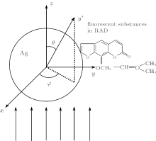
As shown in Fig. 1, we consider a system of a silver sphere with a RAD attached to it. The silver sphere has a radius r and is placed at the origin of a coordinate system. The incident plane wave polarizes in the x direction and propagates along the z direction. We assume that only the dipole oriented along the x axis is excited.[16]
 | Fig. 1. Sketch of the system. |
Firstly, we use a sphere with a radius of 21 nm, whose LSPR band peak is close to the excitation wavelength 315 nm of RAD. For a weak excitation, such as RAD, the excited rate for a two-level system is proportional to the absolute square of the exciting electric field E.[17] The electric field of the incident wave can be expressed as

where k = 2π /λ . The interaction between the incident field and the silver sphere creates a scattered field Es, which can be calculated by the Mie theory.[18, 19] The local field can be expressed as

The RAD is excited by the local field. The excitation enhancement factor can then be defined as

The averaged enhancement factor can be written down as[20– 22]

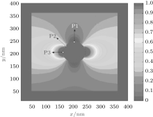
Based on the above theory, we calculate the enhancement factor of the local field around the silver sphere in different orientations by the FDTD method. The field distribution is shown in Fig. 2. The field enhancement regions are concentrated in close vicinity to the surface of the metal sphere, and they can be up to several tens of nanometers.
 | Fig. 2. The local field calculated for the silver sphere (r = 21 nm) via FDTD method. |
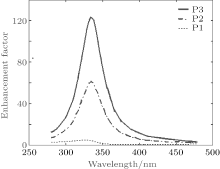
The maximal enhancement factor of the local field can reach up to three orders of magnitude. We exploit the enhancement factors at θ = π /2 and φ = 0, π , which are shown as points P1, P2, and P3 in Fig. 2. Point P1 is placed perpendicularly to the polarization direction, which is the orientation that has the maximum enhancement factor, Point P2 is placed at an angle of 45° relative to the polarization direction, and point P3 is along the polarization direction. All of these three points are in the xy plane. Figure 3 shows the field enhancement factors of points P1, P2, and P3. The enhancement factor is maximum at point P3, which can be up to nearly 120. The enhancement factor is nearly 60 at point P2 and is less than 10 at point P1. The plasmonic near field is distributed with a maximum along the illumination light polarization, which results in the different enhancement of the local field at the three different points.
 | Fig. 3. The enhancement factors at the three points P1, P2, and P3. |
Figure 4 shows the distance-dependent electric field enhancement factor calculated by the Mie theory, and the data are taken along the polarization axis. As shown in Fig. 4, the average electric field enhancement around the Ag nanoparticle decreases quickly as the distance increases. When the distance between the RAD and the sphere is about 20 nm, the field enhancement factor can reach to 125, and it then decreases as the distance increases. The enhancement factor is only several when the distance is larger than 50 nm. This trend of the local field enhancement means that the plasmon resonance decays exponentially. We believe that the plasmonic effect on the luminous enhancement in the near-field region is more pronounced than that in the far-field region.[23]
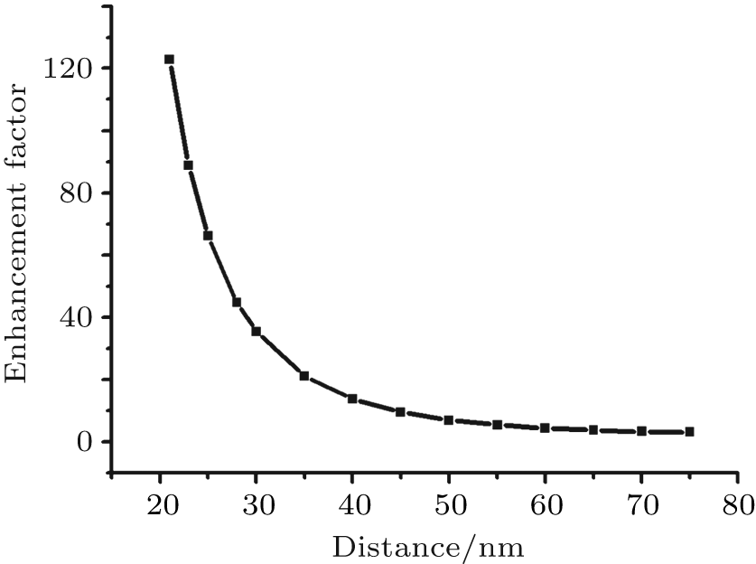 | Fig. 4. The excitation enhancement factor versus the distance away from the center of a 21 nm silver sphere. |
We then consider the situation that the LSPR band of the sphere coincides with RAD’ s radiation wavelength. We use a sphere with a radius of 38 nm, whose LSPR band centers at the emission wavelength of 400 nm of RAD. The LSPR introduces an inhomogeneous environment for the oscillating dipole. Figure 5 shows the extinction spectrum of the silver sphere. For the silver sphere, the quadrupole resonance occurs near 310 nm. The dipole plasmon resonance peaks shifts to near 400 nm, which coincides with RAD’ s emission wavelength. In the vicinity of the silver sphere, the RAD at the excited state can undergo nonradiative decay, emit a photon directly to the far field, or relax rapidly by exciting the LSPR of the sphere via energy transfer. The LSPR can alter both the radiative and the nonradiative decay rates owing to the localized photonic states introduced by the plasmon resonance. Once the LSPR is excited, it can either decay nonradiatively due to internal damping or reradiate into the far field at the emission wavelength.[9, 22]
 | Fig. 5. The extinction spectrum of the silver sphere (r = 38 nm) obtained with the Mie theory. |
As reported in Refs. [15] and [18], the enhancement factor of the radiative rate is

and the energy transfer factor is

where γ 0 is the free emission decay, P0 is the radiation power of the isolated dipole, the total decay rate γ can be divided into a radiative part and a non-radiative part, γ = γ r + γ nr, Pr is the radiation power to the far-field, Pabs is the radiation power absorbed by the environment, and gr(ω ) and gET(ω ) are related to the corresponding cross sections. The emission enhancement factor gem(ω ) can be given as[18, 21]
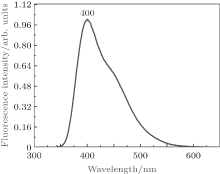
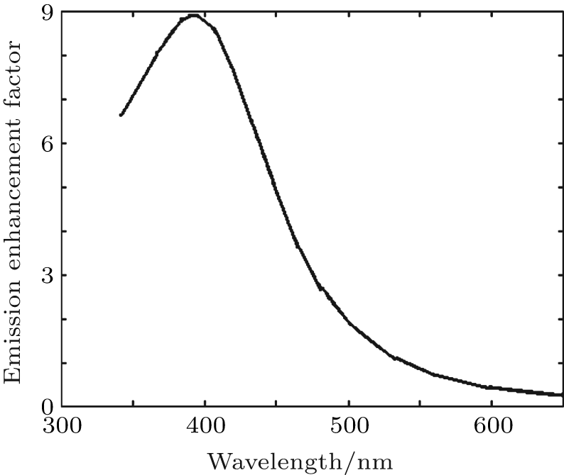
where f0(ω ) is the integral-normalized emission spectrum of RAD in aqueous solution, and the measured emission spectrum is shown in Fig. 6, the emission peak of RAD is at 400 nm. The η 0 is the quantum efficiency of the isolated molecule, which is η 0 ≈ 0.15 for RAD. The fluorescence emission spectrum of RAD is obtained as

 | Fig. 6. The amplitude-normalized emission spectrum of RAD in aqueous solution. |
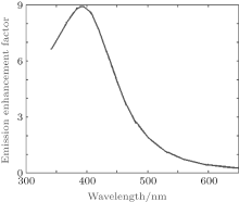
Figure 7 shows the emission enhancement factor calculated for RAD in the vicinity of the single silver sphere (r = 38 nm). As shown in Fig. 7, the enhancement at 400 nm is maximum due to the spectrum overlap between the LSPR band and the emission band of RAD, the largest emission enhancement can reach up to 9 times. The single silver sphere whose resonance wavelength coincides with the emission wavelength enhances the fluorescence emission, however, the enhancement is limited. If the quantum efficiency is too high, then the fluorescence emission would even be quenched.[17, 24]
 | Fig. 7. The emission enhancement factor for RAD in the vicinity of the single silver sphere (r = 38 nm). |
For the excitation process, the incident plane wave interacts with the silver sphere, and the plane wave does not change with the distance. For the emission process, the interaction is between the near field of the dipole and the silver sphere. Unlike the plane wave, the dipole field decreases quickly as the distance becomes longer, this is why the excitation enhancement is greater than the emission enhancement.
In summary, we study the influence of a single silver sphere on the fluorescence properties of RAD. The radius of the sphere is tuned to make its plasmon resonance band coincide with RAD’ s excitation and emission bands, respectively. Based on the numerical calculations, we predict that the excitation enhancement caused by a 21 nm silver sphere is more pronounced than the emission enhancement caused by a 38 nm silver sphere, and the optimum enhancement is more than one hundred times for the luminescence intensity of fluorescence. These results point out the optimal means of enhancing fluorescence in further work.
| 1 |
|
| 2 |
|
| 3 |
|
| 4 |
|
| 5 |
|
| 6 |
|
| 7 |
|
| 8 |
|
| 9 |
|
| 10 |
|
| 11 |
|
| 12 |
|
| 13 |
|
| 14 |
|
| 15 |
|
| 16 |
|
| 17 |
|
| 18 |
|
| 19 |
|
| 20 |
|
| 21 |
|
| 22 |
|
| 23 |
|
| 24 |
|