Self-assembled superparamagnetic nanoparticles as MRI contrast agents— A review
Su Hong-Ying†‡a)  , Wu Chang-Qiang†
b) , Li Dan-Yangc) , Ai Hua§b)
, Wu Chang-Qiang† 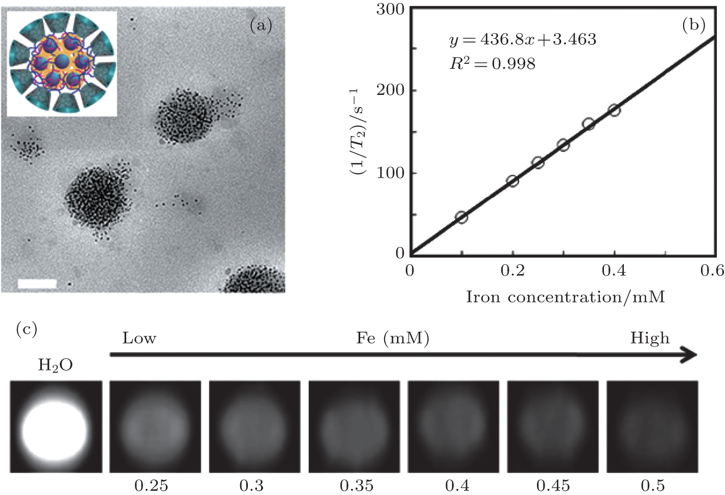
Self-assembled superparamagnetic nanoparticles as MRI contrast agents— A review |
|
Su Hong-Ying†‡
, Wu Chang-Qiang† |
| (a) TEM bright field image of the β–CD-Dex- g -SA/SPIO nanocomposite dried on a formvar coated copper grid (scale bar = 100 nm) and a cross-sectional schematic; (b) T 2 relaxation rate (1/ T 2, s−1) as a function of Fe concentration (mM) for SPIO nanocrystal loaded β–CD-Dex- g -SA micelles at 1.5 T; (c) T 2-weighted MRI images (1.5 T, spin-echo sequence: TR = 5000 ms, TE = 12 ms) of β–CD-Dex- g -SA/SPIO micelles in water. Reprinted with permission from Ref. [ 20 ]. Copyright 2013, Elsevier Ltd. |
| |