Flexible reduced field of view magnetic resonance imaging based on single-shot spatiotemporally encoded technique
Li Jinga) , Cai Cong-Bob) , Chen Lina) , Chen Yingc) , Qu Xiao-Boa) , Cai Shu-Hui†a) 
Flexible reduced field of view magnetic resonance imaging based on single-shot spatiotemporally encoded technique |
|
Li Jing
|
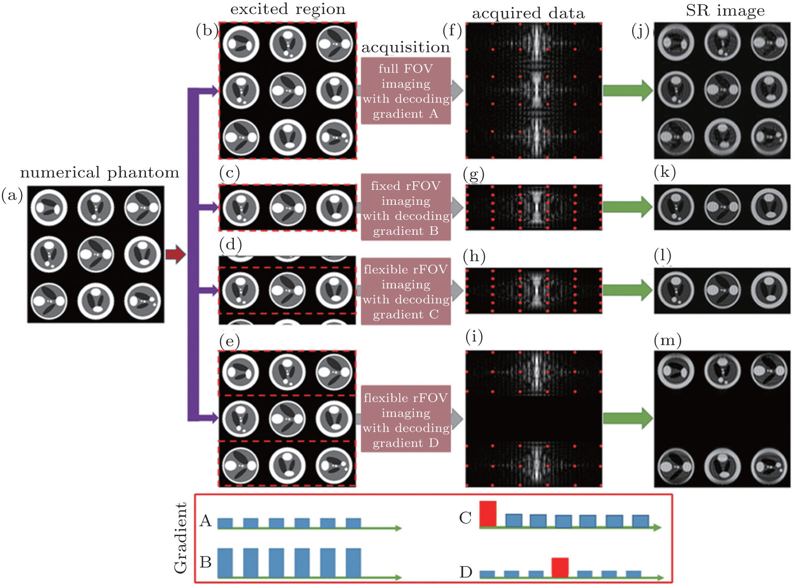
| Schematic diagram of the flexible rFOV imaging method. (a) Numerical phantom. (b) The excited region in full FOV SPEN MRI. (c) The excited region in fixed rFOV SPEN MRI. (d) The excited region in flexible rFOV SPEN MRI with one ROI. (e) The excited region in flexible rFOV SPEN MRI with two ROIs. (f) Full FOV SPEN data obtained with decoding gradient A. (g) Fixed rFOV SPEN data obtained with decoding B. (h) Flexible rFOV SPEN data obtained with decoding gradient C. (i) Flexible rFOV SPEN data obtained with decoding gradient D. (j)–(m) SPEN images after SR reconstruction to panels (f)–(i). Common parameters: Δ O Hz = 64 kHz, T exc = 4 ms, full FOV = 6.0 cm × 6.0 cm, reduced FOV = 6.0 cm × 2.0 cm, and sampling data matrix size = 96 × 96. |
| |