Flexible reduced field of view magnetic resonance imaging based on single-shot spatiotemporally encoded technique
Li Jinga) , Cai Cong-Bob) , Chen Lina) , Chen Yingc) , Qu Xiao-Boa) , Cai Shu-Hui†a) 
Flexible reduced field of view magnetic resonance imaging based on single-shot spatiotemporally encoded technique |
|
Li Jing
|
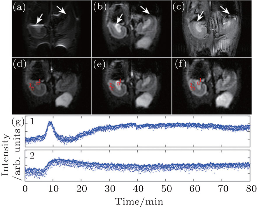
| DCE MR images of rat kidneys with injected contrast agent PL-DF-DOTA-Gd. (a) rFOV EPI image. (b) rFOV SPEN image. (c) Reference multi-shot gradient echo image. (d) rFOV SPEN image that is acquired after the steady-state magnetization condition has been reached and before the contrast agent is injected. (e) rFOV SPEN image acquired near the time when the largest contrast is reached. (f) Final DCE image. (g) DCE time courses of the pixels marked on the images ((d) − (f)). The time courses show the regional variation of the PL-DF-DOTA-Gd effect on the signal of medulla (1, upper) and cortex (2, bottom). |
| |