Flexible reduced field of view magnetic resonance imaging based on single-shot spatiotemporally encoded technique
Li Jinga) , Cai Cong-Bob) , Chen Lina) , Chen Yingc) , Qu Xiao-Boa) , Cai Shu-Hui†a) 
Flexible reduced field of view magnetic resonance imaging based on single-shot spatiotemporally encoded technique |
|
Li Jing
|
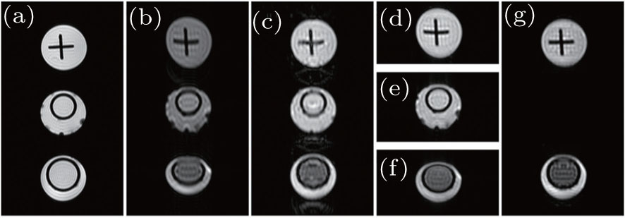
| Imaging results of water phantom. Slice thickness = 2 mm, imaging matrix size = 256 × 256, L x = 40 mm (horizontal), L y = 70 mm [(a)–(c)], 23 mm [(d)–(f)], 46 mm (g) (vertical). (a) Reference multi-scan gradient echo image (sampling data matrix size = 256 × 256, dummy scan = 32, TR = 600 ms, sequence execution time = 172.8 s). (b) Full FOV EPI image (sampling data matrix size = 64 × 64, G acq = 0.0168 T/m, sequence execution time = 47.6 ms). (c) Full FOV SPEN image ( P = 1, G acq = 0.0756 T/m). (d)–(f) The rFOV image obtained with flexible decoding scheme for different ROI ( P ≈ 0.3, G acq = 0.0252 T/m). (g) The rFOV image obtained with flexible decoding scheme for two ROIs ( P ≈ 0.7, G acq = 0.0504 T/m). Common parameters for SPEN imaging: T exc = 3 ms, Δ O Hz = 96 kHz, sampling data matrix size = 64 × 64, R = 32 kHz/ms, sequence execution time = 49.1 ms, and acquisition bandwidth along readout dimension = 250 kHz. |
| |